Other Components Of The Knee
The knee also contains menisci and cartilage which function as shock absorbers, allowing the knee to move with less friction.
- Articular cartilage: is the smooth lining covering the end of the bone. If its damaged or worn away, an individual may develop arthritis of the knee.
- Meniscus: is contained in the area between the end of the thigh bone and the top of the shin bone. It acts as a shock absorber. The meniscus help distribute the bodys weight evenly, adding balance and stability.
Meanwhile, joint bursa found within the knees tissue, is a closed fluid filled sac in front of the knee, just below the surface of the skin. It cushions the bones, tendons, muscles, i.e., smooths out motion between the skin and bone, with the fluid allowing muscles and tendons to glide over ligaments and/or bone.
There are various bursae in the knee, performing this function, including the pre-patellar bursa. And there are hundreds of bursae throughout the body, each acting in a similar capacity for various parts of the anatomy.
Diseases And Afflictions Of The Knee Joint
Falls or accidents often cause injuries to the knee joint. These can occur singly, but also in combination.
- Meniscus lesion
- Inner ligament tear
- Patellar dislocation
The application of strong force, for example an impact with a hard surface, can cause fractures of the bony components of the knee, such as a patella fracture.
Wear and inflammation in the knee can lead to pain. A few examples:
Osteokinematics And Range Of Motion
The ligaments and menisci provide static stability and the muscles and tendons dynamic stability.
The main movement of the knee is flexion – extension. For that matter, knee act as a hinge joint, whereby the articular surfaces of the femur roll and glide over the tibial surface. During flexion and extension, tibia and patella act as one structure in relation to the femur. The quadriceps muscle group is made up of four different individual muscles. They join together forming one single tendon which inserts into the anterior tibial tuberosity. embedded in the tendon is the patella, a triangular sesamoid bone and its function is to increase the efficiency of the quadriceps contractions. Contraction of the quadriceps pulls the patella upwards and extends the knee.Range of motion: extension 0o. The hamstring muscle group consists of the biceps femoris, semitendinosus and semimembranosus. They are situated at the back of the thigh and their function is flexing or bending the knee as well as providing stability on either side of the joint line.Range of motion: flexion 140o.
Secondary movement is internal – external rotation of the tibia in relation to the femur, but it is possible only when the knee is flexed.
Recommended Reading: How To Combat Arthritis In The Knees
Functional Anatomy Of Knee Joint
The specific design of knee joint anatomy allows a number of functions:
The Bones Of The Knee

The knee joint is made up of three bones:
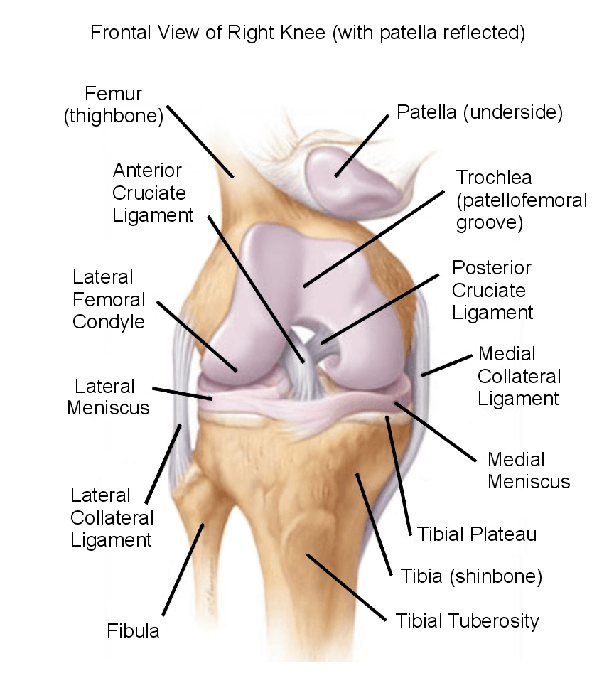
There are two rounded joint surfaces, known as condyles, at the lower end of the femur . The cruciate ligaments run through the gap between these two projections.
The femurs condyles are located opposite two relatively flat, slightly pan-shaped joint surfaces on the tibia . There are two small bumps between them, to which the cruciate ligaments are attached.
The kneecap is located in front of the femur, above the condyles. It is connected to the tibia by the patellar tendon. When the knee is bent or stretched, the slightly wedge-shaped inner side of the kneecap, which is covered with articular cartilage, slides along a groove in the femur. The kneecap reduces the friction between the tendon and the bone, and it keeps the tendon from slipping sideways too. It also extends the lever effect of the femur, improving the transfer of force.
You May Like: What Do You Do For Water On The Knee
Joint Capsule And Lining
The synovium is the lining of the joint space. The synovium is a layer of tissue that defines the joint space.
The synovial cells produce a slippery, viscous fluid called synovial fluid within the joint. In conditions that cause inflammation of the joint, there can be an abundance of synovial fluid produced, which leads to swelling of the knee joint.
Anterior Cruciate Ligament Injury
The anterior cruciate ligament is the most commonly injured ligament of the knee. The injury is common during sports. Twisting of the knee is a common cause of over-stretching or tearing the ACL. When the ACL is injured a popping sound may be heard, and the leg may suddenly give out. Besides swelling and pain, walking may be painful and the knee will feel unstable. Minor tears of the anterior cruciate ligament may heal over time, but a torn ACL requires surgery. After surgery, recovery is prolonged and low impact exercises are recommended to strengthen the joint.
Recommended Reading: What Is The Best Knee Replacement On The Market
A Rather Special Bone: The Kneecap The Largest Human Sesamoid Bone
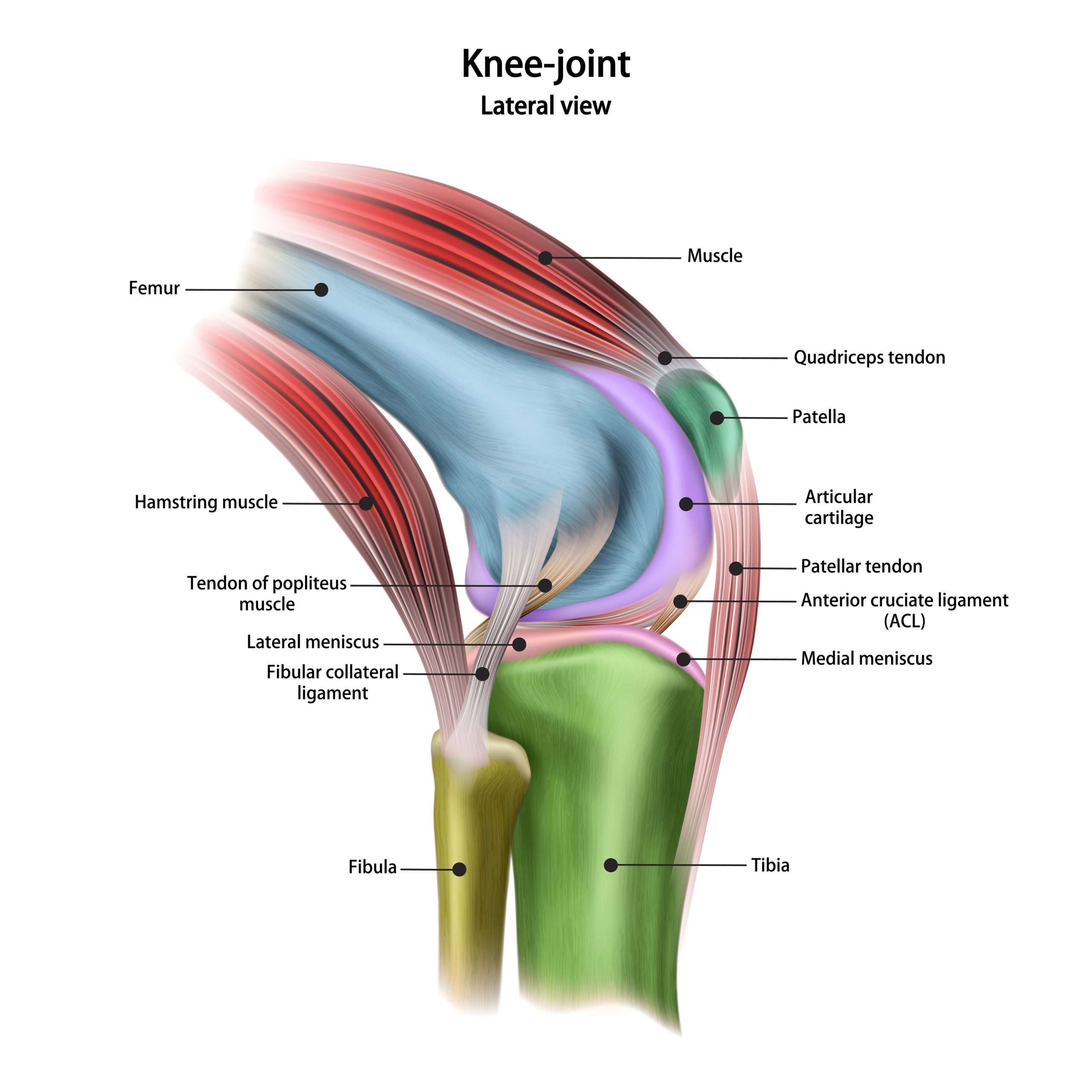
The kneecap also known as the patella is the largest human sesamoid bone. It is fused with the tendon structure that runs from the thigh muscle to the shin bone. The section above the kneecap is called the quadriceps tendon, and the section below the kneecap is called the patellar tendon.
In a sense, the patella acts as a “spacer” between the tendon and the joint, allowing greater leverage and less effort to extend the knee. Moreover, the patella like all sesamoid bones prevents the tendons from being damaged by compressive stress when moving back and forth over the joints.
If we had no kneecaps, incidentally, our thigh muscles would probably be much bigger and we would probably not be able to walk.
Anatomy Of The Knee Bones And Joints Of The Knee Anatomy
The anatomy of the knee consists of 3 main bones:
Lateral Knee Anatomy
The femur and the tibia are the main movers of the joint to allow for the hinge motion. This connection of the femur and tibia is a joint called the tibiofemoral joint. The patella sits on top of the tibiofemoral joint in a groove in the front of the femur. The patella is a floating bone that works as a fulcrum for the quadriceps muscle to function properly. This joint is called the patellofemoral joint and allows the patella to move up and down, and the knee bends and straightens.
Also Check: Why Are My Knees Hot
The Human Knee: Anatomy Motion And How It Works
The knee is one of the largest and most important joints in the human body, tasked with the important job of connecting the upper leg with the lower leg to facilitate motion. At its most basic definition, one may consider the knee to simply be two leg bones joined together by muscles, ligaments, and tendons. But the knee is much more than the sum of its parts. Indeed, its the precise, nimble, effortless way that the joints composition works in unison to create movement that makes the knee such a crucial component of the human anatomy.
So, lets start at the beginning. When talking about the knee, one should start by learning the basic components of the joint.
The knee is composed of three main structures:
- The tibia is the shin bone, the largest bone of the lower part of the leg
- The femur is the thigh bone, part of the upper leg bone
- The patella is also referred to as the kneecap
Lower Leg & Knee Joint Anatomy
There are eight different components of knee joint anatomy:
A really important part of knee joint anatomy is the cartilage. There are two different types of knee cartilage:
- Articular Cartilage: a thin layer of cartilage whichlines the surfaces of the knee joints
- Knee Meniscus: a special extra thick layer of cartilage that sits on the top of the tibia
The knee meniscus is particularly important as it acts as a shock absorber to reduce theforces going through the bones and reduces friction, allowing the bonesto move smoothly.
The back of the patella is also lined with cartilage, the thickest in the whole body due to the immense forces that go through the kneecap.
You can find out all about the different types of knee cartilage, how they work, and cartilage injuries and how to treat them in the knee joint cartilage anatomy section.
Recommended Reading: What To Do For Gout In Your Knee
Synovial Bursa In The Knee: Cushioning For Bones Muscles And Tendons
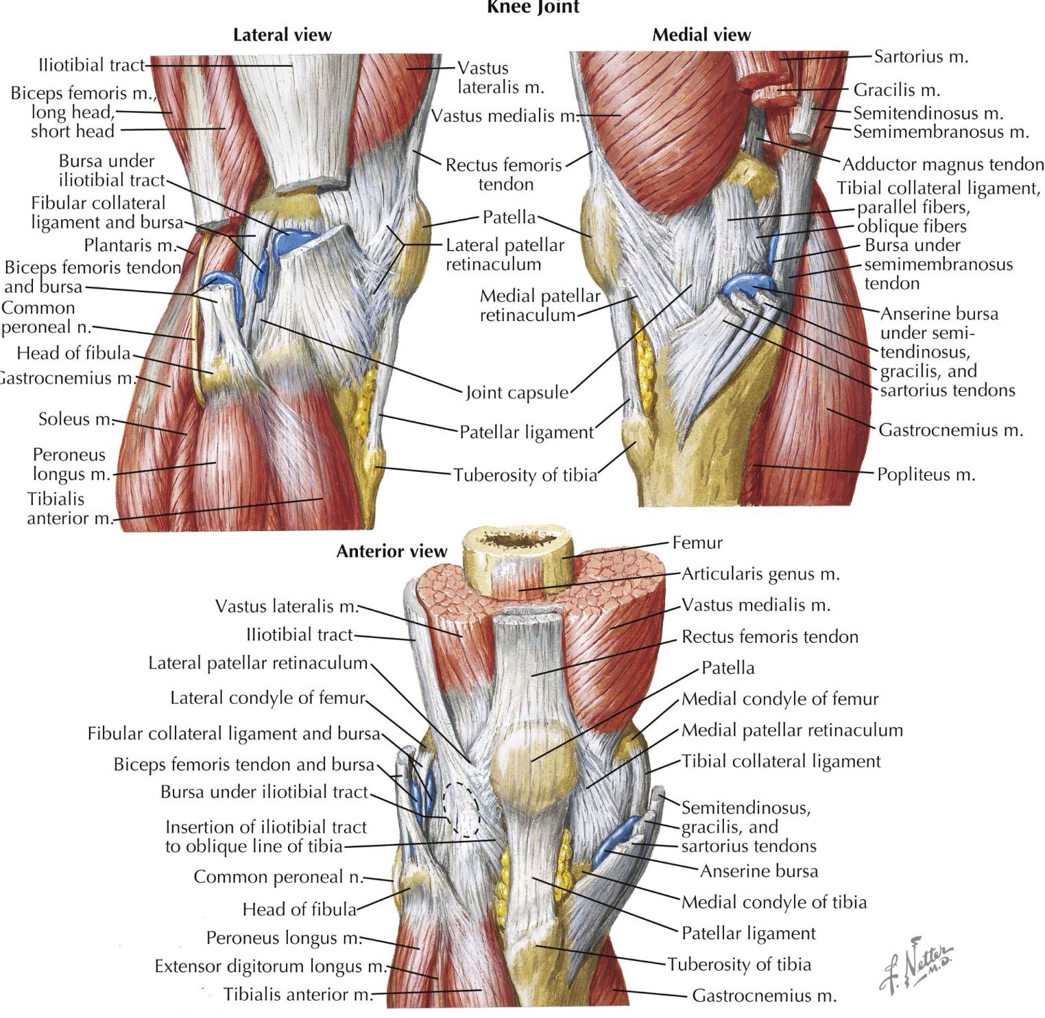
The knee also contains bursae that protect the joint from rubbing and from pressure during movements of the knee. They are found above the kneecap, in front of and behind the patellar tendon. Bursae are slidable, elastic, fluid-filled “pads” that can become inflamed. An inflamed bursa or bursitis can be very painful.
Supporting The Knee Joint: Ligaments Tendons And Muscles

The knee joint is held in place by a prominent ligamentous apparatus. The ligaments prevent the bony structures from rubbing against each other too much.
- Collateral ligaments: The collateral ligaments stabilise the knee mainly against bending stresses in the frontal plane. The ligaments therefore prevent the knee from twisting sideways.
- Cruciate ligaments: The anterior and posterior cruciate ligaments are found inside the knee joint. These two ligaments stabilise the knee against various movements of the tibia in the sagittal plane: The anterior cruciate ligament prevents the tibia from sliding forward . Similarly, the posterior cruciate ligament prevents the tibia from sliding backwards .
Also Check: What Do You Do For Arthritis In The Knee
What Is The Anatomy Of The Knee
The knee joint is made up of bones, muscles, tendons, ligaments, and cartilage. Each individual piece of the complex structure works with the others to provide both fluid movement and stability in the knee. The knee executes many functions including helping the body stay in an upright position, moving to lower and raise the body and also helping to propel the body forward.
The femur , the tibia/fibula and the patella meet to form the knee joint. Muscles are attached to the patella bone through connecting tendons. The quadriceps muscles provide the ability to extend the knee and are connected through the quadricep tendon. The hamstring tendons provide the ability to flex the knee. Tendons transmit muscle forces to the joint. Fibrous bands called ligaments serve as stabilizers during joint movement.
The four major ligaments that connect the upper and lower bones of the knee are:
- Anterior Cruciate Ligament
- Medial Collateral Ligament ,
- Lateral Collateral Ligament .
- Ligaments are tough, fibrous tissues. The cruciate ligaments support the knee in the side plane and limit forward and backward motion of the knee bone. The collateral ligaments support the knee in the frontal plane and limit side to side motion of the joint. All four of these elements of the knee anatomy work collectively to stabilize and support the knee joint. The ligaments prevent any excessive forward or backward sliding motion of the bones within the joint.
Ligaments Of The Knee
Ligaments are structures that connect two bones together. There are four major ligaments that surround the knee joint.
Two of these ligaments are in the center of the joint, and they cross each other. These are called the cruciate ligaments and consist of the anterior cruciate ligament and the posterior cruciate ligament.
One ligament is on each side of the knee jointthe medial collateral ligament on the inner side, and the lateral collateral ligament on the outer side. Ligament injuries typically result in complaints of the instability of the knee joint.
You May Like: Who To See For Knee Pain
What Type Of Joint Is The Knee
The knee is what is known as a synovial hinge joint, but what does that mean?
- Synovial Joint: has a joint capsule which is like a sac surrounding the joint. The capsule contains synovial fluid which nourishes and lubricates the joint allowing it to move smoothly and painlessly – a bit like the oil in your car
- Hinge Joint: typically allows motion in one plane, flexion and extension. The knee is the largest hinge joint in the body and is slightly unusual as it also allows a small amount of rotation.
Normal Anatomy Of The Knee Joint
The knee is made up of four bones. The femur or thighbone is the bone connecting the hip to the knee. The tibia or shinbone connects the knee to the ankle. The patella is the small bone in front of the knee and rides on the knee joint as the knee bends. The fibula is a shorter and thinner bone running parallel to the tibia on its outside. The joint acts like a hinge but with some rotation.
The knee is a synovial joint, which means it is lined by synovium. The synovium produces fluid lubricating and nourishing the inside of the joint. Articular cartilage is the smooth surfaces at the end of the femur and tibia. It is the damage to this surface which causes arthritis.
You May Like: How Do Knee Braces Help Knee Pain
Ligaments & Joint Capsule
The joint capsule has thick and fibrous layer superficially and thinner layers deeper. This along side the capsule ligaments enhances she stability of the knee. As with all of the structures that from the knee they are under most tension therefore more stable in an extended position in comparison to the laxity present in a flexed position . Inside this capsule is a specialized membrane known as the synovial membrane which provides nourishment to all the surrounding structures. The synovial membrane produces synovial fluid which lubricates the knee joint. Other structures include the infrapatellar fat pad and bursa which function as cushions to exterior forces on the knee. The synovial fluid which lubricates the knee joint is pushed anteriorly when the knee is in extension, posteriorly when the knee is flexed and in the semi flexed knee the fluid is under the least tension therefor being the most comfortable position if there is a joint effusion.
The ligaments of the knee maintain the stability of the knee. Each ligament has a particular function in helping to maintain optimal knee stability.
|
|
|
Superior gluteal |
|
S Of A Human Knee Joint Puzzle And Nomenclature
Health Science 3-6, 6-9
Traditionally, the Montessori curriculum introduces human anatomy in the late elementary years in the form of nomenclature cards. At Alisons Montessori, we have been striving to create materials to introduce human anatomy to younger children. This allows them to gain awareness of how their body functions, which empowers them to make healthy choices.
- Wooden Puzzle and Labels
- Anatomy of a Human Knee Joint Complete Set
We would like to demonstrate this fact by presenting our new puzzle, Parts of a Human Knee Joint. The knee joint is an intricate junction in the human body. It is also one of the most complex and stressed joints in the body. Daily activities such as walking, running, and climbing cause shock to the knees, which is absorbed by the cartilage within them. By learning how joints are structured, children will understand why good posture and an active lifestyle is important for proper joint health.
Children will also learn the scientific terms of the bones connected to the joints. The tibia, often called the shinbone, is a thick bone that supports the majority of the bodys weight. It is supported by a thinner bone, the fibula. The femur bone, known as the thigh bone, sits on top of the knee. It is the longest, largest, and heaviest bone in the body. The primary function of the femur is weight bearing and gait stability.
Also Check: How To Treat Arthritis In Knee At Home
Knee Anatomy Joint Capsule
Another important part of knee anatomy is the joint capsule. This is like a bag that surrounds the joint containing synovial fluid to nourishand lubricate the knee allowing it to move smoothly and freely.
Any swelling that occurs in the joint is contained inside the capsule, which is why injuries can cause the knee to “balloon”. If the joint capsule is damaged, swelling is no longer confined to the joint so tends to actually be less obvious.
You can find out more in the knee swelling section.
The Muscles In The Back Of The Knee

Hamstring Muscle diagram
- Gastrocs: A group of 2 muscles that sit in on the lower leg backside that works in tandem with the hamstrings to cause the knee to bend. The gastroc or calf muscle can be strained and torn during sports like tennis or basketball. The athlete will feel a “pop’ in the calf.
Calf Muscle Diagram
- Tendons attach the knee muscles to the bone. The two patellar tendons can also be prone to overuse and the development of patellar tendonitis. Jumper’s knee is common in the knee with athletics.
All of these muscles also have functions at different joints such as the hip and the ankle. Injuries to these structures, such as a pull or strain, will cause pain when activating the muscle and, if severe enough, will cause significant weakness.
Also Check: Can Arthritis In Knee Be Cured