What Are The Grades Of Meniscus Tears
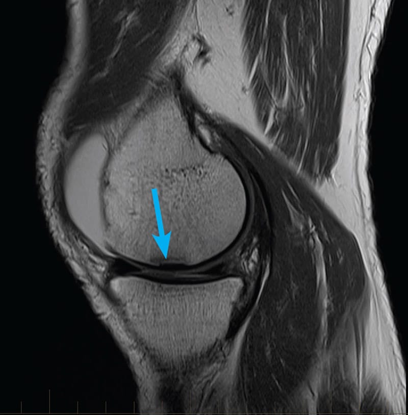
Grade 0, normal intact meniscus Grade I, intrasubstance globular-appearing signal not extending to the articular surface Grade II, linear increased signal patterns not extending to the articular surface Grade III, abnormal signal intersects the superior and/or inferior articular surface of the meniscus, an
Sizes Shapes And Patterns
Multiple cross-sectional representations of a meniscal tear have to be translated into a 3-dimensional description for the benefit of the arthroscopist.
Meniscal tears occur in 2 primary planes: vertical and horizontal. The 3 basic meniscal tear shapes are longitudinal, radial, and horizontal. Meniscal tears are either partial thickness or full thickness.
Vertical tears are aligned perpendicular to the coronal plane of the meniscus and can be subdivided into longitudinal and radial tears. They occur as traumatic lesions in younger patients. A full-thickness vertical tear contacts both the superior and inferior articular meniscal surfaces, completely dividing the torn part of the meniscus into an inner and outer portion. Such tears can lead to the development of bucket-handle tears.
Longitudinal tears separate the meniscus into inner and outer fragments and occur parallel to the outer margin of the meniscus . These tears are equidistant from the outer meniscal margin throughout their entire course.
Short tears, or those confined to the posterior horn, may be visible only on sagittal images. Longer tears propagate into the body of the meniscus. These are seen on both sagittal and coronal images.
Bucket-handle tears
Radial tears
Radial tears are vertical tears and propagate perpendicular to the main axis of the meniscus. These injuries are devastating because a full-thickness tear destroys meniscal integrity .
Horizontal tears
Miscellaneous tears
The History Of Mri Findings And Knee Pain
You would think that if your doctor sees a problem like a meniscus tear on your MRI that this would be the cause of your knee pain. After all, something or nothing happened and your knee began to hurt. Hence, it makes sense that the cause of that pain could be seen on an MRI scan. However, the hard truth is more complex.
Back in 2008 researchers publishing in the New England Journal of Medicine reported that most people over age 35 without knee pain had everything from meniscus tears to cartilage loss on their MRIs . These findings began the first major questioning of whether meniscus tear MRI had much value. Meaning, were meniscus tears a problem or just a function of normal aging like wrinkles?
As more research came together, Scandinavian scientists in 2014 published an editorial that physicians and patients alike should look at meniscus tears with the same diagnostic weight we give wrinkles . Again, that meniscus tears were about as important as wrinkles on your face, they are simply of a sign of normal aging.
Do you want to learn more about how to read your knee MRI? See my video below:
Also Check: Why Does My Knee Hurt When I Kneel On It
Indirect Signs Of Meniscal Tears
Indirect signs of meniscal tears include meniscal and parameniscal cysts, joint effusion, medial collateral ligament edema, and bone marrow edema. Bone marrow edema can be 1 of 3 types: perivascular edema occurs around vascular channels and does not extend to the joint surface, linear subchondral marrow edema occurs adjacent to the meniscus and probably reflects hyperemia and increased fluid at the cortical-chondral-meniscal interface, and nonlinear subchondral marrow edema is associated with abnormalities at the adjacent cartilage surface. The presence of this type of edema reflects cartilage loss, chronicity, and extent of meniscal tear.
Protocols And Imaging Views

The knee is generally extended in slight external rotation to facilitate imaging of the anterior cruciate ligament . High spatial resolution is necessary to show meniscal tears. This typically requires a field of view of 14cm × 16cm. For this review, we use 0.4mm × 0.4mm resolution for proton-density-weighted images and 0.5mm × 0.5mm resolution for fat sat T2-weighted images. An extremity coil optimizes the signal-to-noise ratio .
Images should be obtained on all three planes: sagittal, coronal, and axial. Sagittal images are obtained with the knee in slight external rotation to visualize the anterior cruciate ligament . Several factors should be taken into account to optimize the imaging protocols. Although imaging on all three planes is useful, all sequences should not be performed on all planes. Usually T1-weighted sequences are performed on the sagittal plane while T2-weighted sequences are performed on all three spatial planes .
Sequences defining anatomical structures should be distinguished from those characterizing meniscal pathologies. Imaging of meniscal structures and contours is better with proton-density T2-weighted sequences:
Don’t Miss: My Knee Pops And Hurts
What Causes A Medial Meniscus Injury
The medial meniscus is injured more often than the lateral meniscus due to its location and attachment site within the knee. The medial meniscus is attached to the medial collateral ligament and the joint capsule. It is trapped in an area of vulnerability, especially during forced movements such as a rapid stop, twisting motion when the foot is planted, or from a hard fall or tackle in sports. The medial meniscus also handles a lot of load and load distribution which can wear down the fibrocartilage and cause it to tear or fray. The most common causes of a medial meniscus injury involve sports such as:
Prevalence Of Meniscal Damage
In the overall sample, the prevalence of meniscal damage in the right knee was 35% . Damage to the medial meniscus was more common than damage to the lateral meniscus . The prevalence of a meniscal tear in the total sample was 31% meniscal destruction was present in 10% . After excluding subjects who had had previous knee surgery , the prevalence of meniscal destruction was 8%.
You May Like: What Is Mechanical Knee Pain
Tips To Prevent Meniscus Tears
You can prevent meniscus tears by regularly performing exercises that strengthen your leg muscles. This will help stabilize your knee joint to protect it from injury.
You can also use protective gear during sports or a brace to support your knee during activities that may increase your risk of injury.
Criteria For Meniscal Tears
Two MRI criteria have been established for diagnosing meniscal tears. If prior surgery has not been performed on the meniscus, the accuracy in diagnosing tears is 90%.
Criterion 1
Criterion 1 is increased internal signal intensity in the meniscus. An article discussed the concept of the “Two-Slice-Touch Rule.” The authors described a positive predictive value of 94% for meniscal tears for the medial meniscus and 96% for the lateral meniscus when the tear is present on 2 consecutive images. The positive predictive value was 55% and 36% for medial and lateral meniscal tears, respectively, when seen on only 1 slice.
The abnormal signal intensity must be in contact with 1 articular surface, either the superior or interior surface or at the tip of the meniscus. If the contact with the articular surface appears on 2 or more consecutive images, the accuracy of the diagnosis of meniscal tear increases.
The rate of proton rotation is shortened by the interaction of synovial fluid and large macromolecules, resulting in shortening of T1 and T2 values, increasing the sensitivity of PD-weighted images in revealing meniscal pathology. Such changes cause a local increase in the degree of freedom of trapped water molecules, resulting in increased T2 times, allowing the detection of increased signal with short-TE sequences. Increased signal within the meniscus is best seen on short-TE images obtained by using PD-weighted or GRE sequences.
Criterion 2
Recommended Reading: Does Medicare Cover Knee Braces
Description Of Lesions: Size Shapes And Characteristics
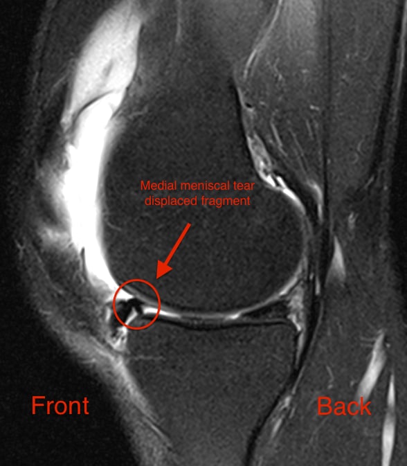
Multiple images of meniscal tears should be translated into 3D images . Meniscal tears occur on two main planes: vertical and horizontal. The three basic shapes of meniscal tears are longitudinal, radial, and horizontal. Meniscal tears are either partial or full thickness .
5.5.1. Vertical Tears
Vertical tears are perpendicular to the coronal plane of the meniscus and can be subdivided into peripheral longitudinal or radial tears. They usually occur following a trauma in young patients .
A vertical tear in the meniscal tissue communicating with the superior and inferior meniscal articular surfaces completely divides the meniscus into two parts .
A vertical tear in the meniscal tissue communicating with the superior and inferior meniscal articular surfaces completely divides the meniscus into two parts. Coronal T2 FSE Fat Sat MRI , Sagittal T2 FSE Fat Sat MRI , Axial T2 FSE Fat Sat MRI , and sagittal T1-weighted sequence MRI. Three-dimensional diagram shows a vertical and longitudinal tear of the meniscus. Three-dimensional diagram shows a vertical tear.
These tears can result in the development of bucket-handle tears . Vertical tears of the posterior horn may not be visible on sagittal images.
5.5.2. Radial Tears
Radial tears are vertical tears that extend perpendicular to the main axis of the meniscus. The most frequent location is the middle segment of the meniscus .
Oblique tears are a type of radial tear .
How Do I Know I Have A Torn Meniscus
Sometimes a torn meniscus can occur and you may not even notice it at first, which doctors may refer to as more of a mild tear. When you experience a mild tear of the meniscus you might notice a slight pain when it happens but really start to notice something is wrong when youve had a chance to rest from your activity and start to notice any pain or swelling around your knee. Even in mild cases, though, the slight pain may linger for a few weeks until the knee heals.
In more moderate to severe cases, it could be quite obvious to you that you have injured your knee because of how much pain you are in. In moderate cases of a torn meniscus, you may even notice that the swelling gets worse the first few days after the injury and you might feel uncomfortable when you try to bend or straighten your knee. Severe cases are rarer but not unheard of, and when this happens you may not even be able to bend your knee and may have a lot of difficulty walking.
Read Also: Knee Replacement Surgery Recovery Time Off Work
Meniscal Tear Patternsmark H Awh Md
A gradient-echo T2*-weighted sagittal image demonstrates a tear within the posterior horn of the medial meniscus . The preferred nomenclature for this tear pattern is:
A gradient-echo T2*-weighted sagittal image
A. Radial Tear
Figure 2:
Although C, a vertical tear, is commonly used to describe such an appearance, the better answer is D, a longitudinal tear. Even better would be to describe a peripheral longitudinal tear extending to the tibial surface within the posterior horn of the medial meniscus!
Although C, a vertical tear, is commonly used to describe such an appearance, the better answer is D, a longitudinal tear. Even better would be to describe a peripheral longitudinal tear extending to the tibial surface within the posterior horn of the medial meniscus!
Medial Collateral Ligament And Posterolateral Corner

The MCL, made up of superficial and deep bands, is the most commonly injured ligament in the knee . Fortunately, the MCL has great potential for healing, and an isolated injury of the MCL can typically be managed conservatively . Although MR is excellent at evaluating the MCL, low-grade injuries are likely overestimated as fluid and edema around the MCL or within the MCL bursa can be seen with multiple other pathologies, including medial meniscal tears and osteoarthritis . This medial compartment pathology can result in bulging of the MCL with fluid deep to the MCL that is reactive and not indicative of a sprain . There is also a subset of patients with deep MCL injuries located typically at the proximal femoral attachment, which may have persistent pain despite conservative therapy and may benefit from surgery . The same pathology that may result in overestimation of low-grade MCL injuries can lead to under diagnosing of deep MCL tears. Another potential problem when evaluating the MCL is injuries at or near the distal tibial attachment. Standard field of view on dedicated MR imaging of the knee often excludes the distal tibial attachment, and edema and fluid around the distal portion of the MCL should raise suspicion for injury and prompt additional imaging .
Fig. 16
A 26-year-old male with partial tear of the MCL. Coronal PD-weighted fat-suppressed images show a tear of the deep MCL fibers along with a low-grade injury of the superficial fibers
Also Check: How Do You Get Rid Of Fluid In Knee
Diagnosing Knee Injury With An Mri
Pinpointing the Cause of Ligament, Tendon, or Meniscus Injury
Magnetic resonance imaging is a technology often used to investigate the sources of knee problems. It works by creating a magnetic field that causes the water molecules in tissue, bones, and organs to orient themselves in different ways. These orientations are then translated into images we can use for diagnosis.
MRIs are not used on their own to make a diagnosis but can often provide strong evidence to support one. When faced with a knee injury, infection, or joint disorder, doctors will often use an MRI to not only pinpoint the cause but to help direct the treatment plan.
While some people find MRIs distressing, either because they are claustrophobic or jarringly noisy, they are invaluable tools which offer a less invasive means of diagnosis.
How Are Medial Meniscus Injuries Diagnosed
Dr. Williams will discuss your symptoms and medical history and will perform a physical examination of the affected knee. He will check for tenderness along the inside of the knee and will perform a few tests to determine range of motion. To confirm his diagnosis, imaging test may be required which can include x-ray, or an MRI scan. MRI stands for Magnetic Resonance Imaging which provides complete views of both the bones and soft tissues in the knee. Most meniscus injuries are seen on an MRI and proper treatment can be planned.
Also Check: Mako Total Knee Replacement Problems
What Causes A Meniscus Tear
A meniscus tear is usually caused by twisting or turning quickly, often with the foot planted while the knee is bent. These tears can occur when you lift something heavy or play sports. Other knee injuries, such as a torn ligament, can happen at the same time. As you get older, your meniscus gets worn. This can make it tear more easily.
Stable Versus Unstable Tears
The stability of tears is determined by a number of factors, including the length, location, and completeness of the tear. Probing the meniscal tear during arthroscopy is critical for determining stability.
A stable vertical longitudinal tear occurs when the central fragment of a meniscal tear cannot be displaced more than 3 mm from the intact meniscal periphery. Any meniscal tear with a displaced fragment is unstable.
Longitudinal tears that are relatively long are unstable their length is assessed on multiple 3- to 4-mm sections in either plane, and they extend through the full thickness of the meniscus or contain fluid on T2-weighted images.
Some meniscal tears do not show a free fragment at the time of MRI. However, it is considered an unstable tear whenever the inner margin of the tear can be displaced to a position where it can be entrapped between the rotating femur and tibia when probed at arthroscopy.
Don’t Miss: What Are The Symptoms Of Knee Pain
Is A Compression Sleeve Good For A Torn Meniscus
Compression sleeves are often the best knee brace for a torn meniscus if you also suffer from arthritic knees or from a degenerative condition. They are also a good choice for an athlete at the end of the rehabilitation process and requiring compression therapy to reduce pain and promote more rapid healing.
Treatment For A Meniscus Tear
Specific treatment for a meniscus tear will be determined by your doctor based on:
-
Your overall health and medical history
-
How bad your injury is
-
How well you can tolerate specific medications, procedures, and therapies
-
The length of time it will take to heal
-
Your opinion or preference
-
Arthroscopic surgery
Also Check: How Long To Recover From Knee Arthroscopy Surgery
Does It Matter Where The Meniscus Tears
The meniscus has two zones, the red zone and the white zone. The red zone is located on the outer third of the meniscus and has a rich blood supply which gives it the ability to heal some tears. The white zone, on the inner two-thirds of the meniscus, does not have a significant blood supply and cannot heal by itself. The location of a meniscus injury is important in determining the plan of treatment. Dr. Williams will diagnose the type of tear and form a treatment plan for his patients in the New York City area that will be the best, individualized option for their specific type of meniscus injury.
What Gets Stored In A Cookie

This site stores nothing other than an automatically generated session ID in the cookie no other information is captured.
In general, only the information that you provide, or the choices you make while visiting a web site, can be stored in a cookie. For example, the site cannot determine your email name unless you choose to type it. Allowing a website to create a cookie does not give that or any other site access to the rest of your computer, and only the site that created the cookie can read it.
Read Also: Can You Overdo It After Knee Surgery
How To Read An Mri Of A Medial Meniscus Tear
Minnesota knee surgeon, Dr. Robert LaPrade details the specifics on how to read an MRI of a medial meniscus tear. There are different types of meniscus tears and a horizontal cleavage tear occurs within the fibers of the meniscus and splits the meniscus in the top and bottom pieces.
To begin, we start with a sagittal view on the lateral side. As we start to go more towards the midline we start to see the lateral meniscus. There is a dark appearance to it, so there is no evidence of disruption. As we scan further we see the ACL and PCL, which both look normal.
Moving more towards the medial side of the knee there is evidence of signal changes in the medial meniscus. In this case, we see a complete white pass of fluid in the meniscus, which indicates that there is a horizontal cleavage tear.
The next view is a coronal scan. As we course more posteriorly we can see the meniscus is in relatively good position, but we are starting to see increase signal in the body of the meniscus, which is indicative of a tear. All the way to the posterior medial aspect we can see signal intensity, which is consistent with the horizontal cleavage tear.
The last view we look at is an axial image. In some cases it is challenging to see the tear within the meniscus from this view, but it is important to assess.