Implant Designs And Sizes
Generic base models of TKR femur components and tibia plates were designed based on widely used commercial products . The base models were scaled to the sizes listed for five manufacturers models as reported in size charts within their respective surgical technique manuals .3). This method was used as access to the official geometry of all the manufacturer models and sizes utilized within the study was not possible. Nor was acquiring physical samples for reverse engineering. Utilizing generic implant shapes also allowed for the test models to be easily controlled/edited for the purposes of the study.
Method for evaluating fit of implant model on subject ground truth anatomy : Generic implant component designs, edges of scaled components, scaled components fitted to subject 3D model predictions, scaled components fitted to subject ground truth anatomy. Dashed box in shows detail not included in fit analysis. Red squares in and show the location of the maximum OUH.
Recognizing The Signs Of A Blood Clot
Follow your orthopaedic surgeon’s instructions carefully to reduce the risk of blood clots developing during the first several weeks of your recovery. They may recommend that you continue taking the blood thinning medication you started in the hospital. Notify your doctor immediately if you develop any of the following warning signs.
Warning signs of blood clots. The warning signs of possible blood clots in your leg include:
- Increasing pain in your calf
- Tenderness or redness above or below your knee
- New or increasing swelling in your calf, ankle, and foot
Warning signs of pulmonary embolism. The warning signs that a blood clot has traveled to your lung include:
- Sudden shortness of breath
- Sudden onset of chest pain
- Localized chest pain with coughing
Types Of Arthritis That Affect The Knee
Inflammatory arthritis
This broad category includes a wide variety of diagnoses including rheumatoid arthritis, lupus, gout and many others. It is important that patients with these conditions be followed by a qualified rheumatologist as there are a number of exciting new treatments that may decrease the symptoms and perhaps even slow the progression of knee joint damage.
Patients with inflammatory arthritis of the knee usually have joint damage in all three compartments and therefore are not good candidates for partial knee replacement. However, inflammatory arthritis patients who decide to have total knee replacement have an extremely high likelihood of success. These patients often experience total, or near-total, pain relief following a well-performed joint replacement.
Osteoarthritis
Osteoarthritis is also called OA or degenerative joint disease. OA patients represent the large majority of arthritis sufferers. OA may affect multiple joints or it may be localized to the involved knee. Activity limitations due to pain are the hallmarks of this disease.
OA patients who have symptoms limited to one compartment of the knee sometimes are good candidates for minimally-invasive partial knee replacement .
Read Also: How To Stop Knee Pain While Sleeping
How Your New Knee Is Different
Improvement of knee motion is a goal of total knee replacement, but restoration of full motion is uncommon. The motion of your knee replacement after surgery can be predicted by the range of motion you have in your knee before surgery. Most patients can expect to be able to almost fully straighten the replaced knee and to bend the knee sufficiently to climb stairs and get in and out of a car. Kneeling is sometimes uncomfortable, but it is not harmful.
Most people feel some numbness in the skin around their incisions. You also may feel some stiffness, particularly with excessive bending activities.
Most people also feel or hear some clicking of the metal and plastic with knee bending or walking. This is normal. These differences often diminish with time and most patients find them to be tolerable when compared with the pain and limited function they experienced prior to surgery.
Your new knee may activate metal detectors required for security in airports and some buildings. Tell the security agent about your knee replacement if the alarm is activated.
Can Rehabilitation Be Done At Home

All patients are given a set of home exercises to do between supervised physical therapy sessions and the home exercises make up an important part of the recovery process. However, supervised therapy–which is best done in an outpatient physical therapy studio–is extremely helpful and those patients who are able to attend outpatient therapy are encouraged to do so.
For patients who are unable to attend outpatient physical therapy, home physical therapy is arranged.
You May Like: Top Of Knee Hurts When Bending
What Is A Knee Replacement Surgery
Knee replacement, also called knee arthroplasty or total knee replacement, is a surgical procedure toresurface a knee damaged by arthritis. Metal and plastic parts are used tocap the ends of the bones that form the knee joint, along with the kneecap.This surgery may be considered for someone who has severe arthritis or asevere knee injury.
Various types of arthritis may affect the knee joint. Osteoarthritis, adegenerative joint disease that affects mostly middle-aged and olderadults, may cause the breakdown of joint cartilage and adjacent bone in theknees. Rheumatoid arthritis, which causes inflammation of the synovialmembrane and results in excessive synovial fluid, can lead to pain andstiffness. Traumatic arthritis, arthritis due to injury, may cause damageto the cartilage of the knee.
The goal of knee replacement surgery is to resurface the parts of the kneejoint that have been damaged and to relieve knee pain that cannot becontrolled by other treatments.
Characteristics Of Severe Arthritis Of The Knee
Pain
Pain is the most noticeable symptom of knee arthritis. In most patients the knee pain gradually gets worse over time but sometimes has more sudden flares where the symptoms get acutely severe. The pain is almost always worsened by weight-bearing and activity. In some patients the knee pain becomes severe enough to limit even routine daily activities.
Stiffness
Morning stiffness is present in certain types of arthritis. Patients with morning stiffness of the knee may notice some improvement in knee flexibility over the course of the day. Rheumatoid arthritis patients may experience more frequent morning stiffness than patients with osteoarthritis.
Swelling and warmth
Patients with arthritis sometimes will notice swelling and warmth of the knee. If the swelling and warmth are excessive and are associated with severe pain, inability to bend the knee, and difficulty with weight-bearing, those signs might represent an infection. Such severe symptoms require immediate medical attention. Joint infection of the knee is discussed below.
Location
The knee joint has three compartments that can be involved with arthritis . Most patients have both symptoms and findings on X-rays that suggest involvement of two or more of these compartments for example, pain on the lateral side and beneath the kneecap . Patients who have arthritis in two or all three compartments, and who decide to get surgery, most often will undergo total knee replacement .
Don’t Miss: Tendons Behind The Knee Hurt
What Is Kinematic Alignment
Movement of the knee joint is achieved through the biomechanical interaction of the soft tissue component together with femoral and tibial articulating surfaces. Mean femoral joint angle is approximately 3° valgus and tibial joint angle measures 3° varus to their respective mechanical axes . Consequently, mean constitutional knee joint alignment is 3° varus to the mechanical axis of the lower limb.
First described by Freeman et al., the MA approach to TKA remains the gold standard. The technique aims to create a neutral lower limb alignment. This is achieved through preparing the distal femoral and proximal tibial cuts perpendicular to their respective mechanical axes. In addition, the posterior femoral condyle is cut in 3° external rotation. The net effect is thought to be equal load distribution through a newly orientated joint line with favourable survivorship outcomes reported when this is achieved .
Appreciation of the KA approach requires an understanding of the kinematic axis of the knee joint. Much of the biomechanical rationale behind KA was introduced by Hollister et al. in the early 1990s and more recently by the work of Eckhoff et al. . Whilst the mechanical axis of the knee is based on a 2D schema of the joint , the kinematic axis refers to its 3D orientation. By definition, the three kinematic axes include:
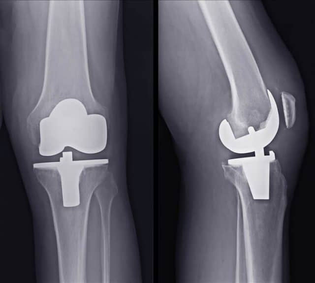
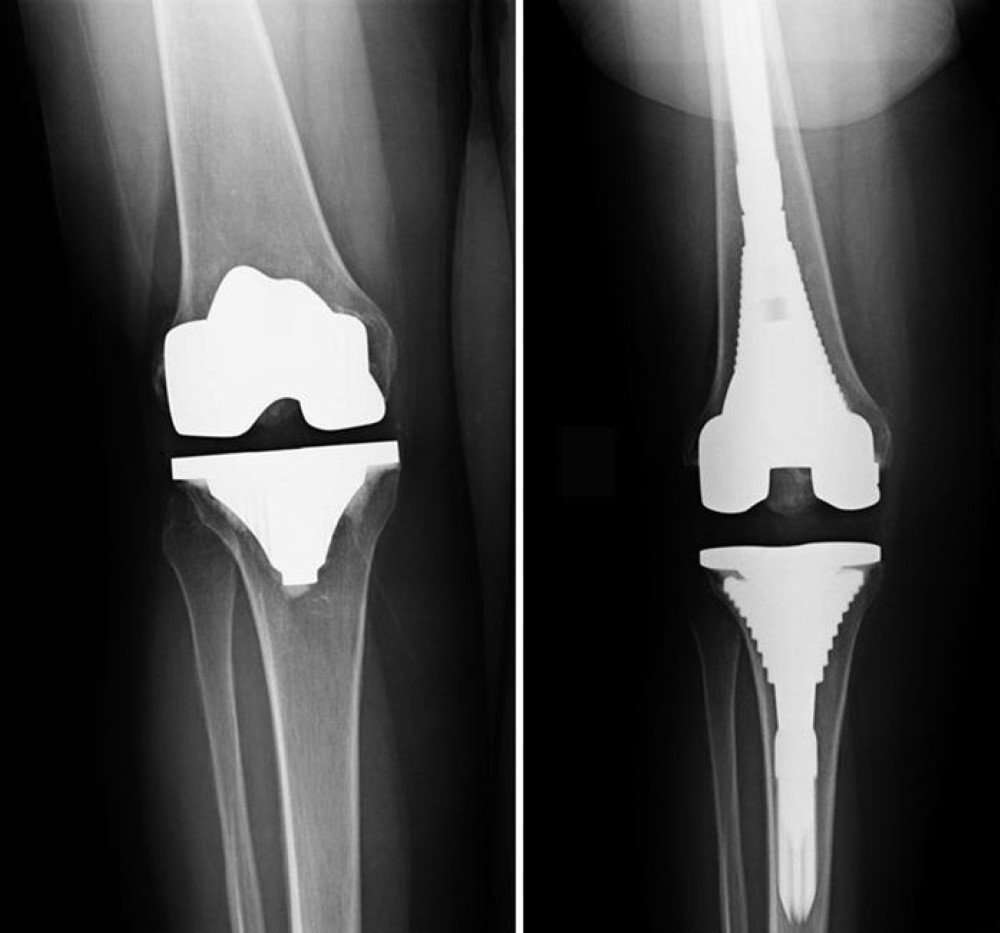
Imaging Of Total Knee Arthroplasty
Chapter 10 Imaging of Total Knee Arthroplasty
Michael K. Brooks, Christopher J. Palestro, Barbara N. Weissman
Total knee arthroplasty results in improvement in overall quality of life in more than 90% of patients.79 This remarkable success and factors such as an aging population have led to a dramatic increase in the number of procedures performed and to corresponding increases in the number of imaging studies obtained for assessment . In addition to radiography, computed tomography , magnetic resonance imaging , ultrasonography , arthrography, and a number of nuclear medicine studies are now available for evaluation of knee arthroplasty. It is the responsibility of both imagers and clinicians to provide the most efficacious and cost-effective examinations in a particular clinical situation. This chapter reviews some available imaging techniques, expected findings in uncomplicated and complicated cases, and the efficacy of various techniques in assessing complications.
Also Check: Can You Get Cancer In Your Knee
When Surgery Is Recommended
There are several reasons why your doctor may recommend knee replacement surgery. People who benefit from total knee replacement often have:
- Severe knee pain or stiffness that limits everyday activities, including walking, climbing stairs, and getting in and out of chairs. It may be hard to walk more than a few blocks without significant pain and it may be necessary to use a cane or walker
- Moderate or severe knee pain while resting, either day or night
- Chronic knee inflammation and swelling that does not improve with rest or medications
- Knee deformity a bowing in or out of the knee
- Failure to substantially improve with other treatments such as anti-inflammatory medications, cortisone injections, lubricating injections, physical therapy, or other surgeries
Total knee replacement may be recommended for patients with bowed knee deformity, like that shown in this clinical photo.
Your Recovery At Home
You may need some help at home for several days to several weeks after discharge. Before your surgery, arrange for a friend, family member or caregiver to provide help at home. You may need a walker, cane, or crutches for the first few days or weeks until you are comfortable enough to walk without assistance.
Read Also: What Is Recovery Like After Knee Replacement Surgery
What Gets Stored In A Cookie
This site stores nothing other than an automatically generated session ID in the cookie no other information is captured.
In general, only the information that you provide, or the choices you make while visiting a web site, can be stored in a cookie. For example, the site cannot determine your email name unless you choose to type it. Allowing a website to create a cookie does not give that or any other site access to the rest of your computer, and only the site that created the cookie can read it.
Risks Of The Procedure

As with any surgical procedure, complications can occur. Some possiblecomplications may include, but are not limited to, the following:
-
Blood clots in the legs or lungs
-
Loosening or wearing out of the prosthesis
-
Continued pain or stiffness
The replacement knee joint may become loose, be dislodged, or may not workthe way it was intended. The joint may have to be replaced again in thefuture.
Nerves or blood vessels in the area of surgery may be injured, resulting inweakness or numbness. The joint pain may not be relieved by surgery.
There may be other risks depending on your specific medical condition. Besure to discuss any concerns with your doctor prior to the procedure.
Read Also: Black Dress With Knee High Boots
Technical Details Of Total Knee Replacement
Total knee replacement surgery begins by performing a sterile preparation of the skin over the knee to prevent infection. This is followed by inflation of a tourniquet to prevent blood loss during the operation.
Next, a well-positioned skin incision–typically 6-7 in length though this varies with the patients size and the complexity of the knee problem–is made down the front of the knee and the knee joint is inspected.
Next, specialized alignment rods and cutting jigs are used to remove enough bone from the end of the femur , the top of the tibia , and the underside of the patella to allow placement of the joint replacement implants. Proper sizing and alignment of the implants, as well as balancing of the knee ligaments, all are critical for normal post-operative function and good pain relief. Again, these steps are complex and considerable experience in total knee replacement is required in order to make sure they are done reliably, case after case. Provisional implant components are placed without bone cement to make sure they fit well against the bones and are well aligned. At this time, good function–including full flexion , extension , and ligament balance–is verified.
Finally, the bone is cleaned using saline solution and the joint replacement components are cemented into place using polymethylmethacrylate bone cement. The surgical incision is closed using stitches and staples.
Anesthetic
Length of total knee replacement surgery
Pain and pain management
Preparation For Total Knee Replacement Surgery
Patients undergoing total knee replacement surgery usually will undergo a pre-operative surgical risk assessment. When necessary, further evaluation will be performed by an internal medicine physician who specializes in pre-operative evaluation and risk-factor modification. Some patients will also be evaluated by an anesthesiologist in advance of the surgery.
Routine blood tests are performed on all pre-operative patients. Chest X-rays and electrocardiograms are obtained in patients who meet certain age and health criteria as well.
Surgeons will often spend time with the patient in advance of the surgery, making certain that all the patient’s questions and concerns, as well as those of the family, are answered.
Costs
The surgeon’s office should provide a reasonable estimate of:
- the surgeon’s fee
- the degree to which these should be covered by the patient’s insurance.
Total Knee Replacement Surgical Team
The total knee requires an experienced orthopedic surgeon and the resources of a large medical center. Some patients have complex medical needs and around surgery often require immediate access to multiple medical and surgical specialties and in-house medical, physical therapy, and social support services.
Finding an experienced surgeon to perform your total knee replacement
Some questions to consider asking your knee surgeon:
- Are you board certified in orthopedic surgery?
- Have you done a fellowship in joint replacement surgery?
- How many knee replacements do you do each year?
Don’t Miss: How To Strengthen Knee Muscles
Condylar Twist Angle 1160
In the kneeling view, it is measured as the angle between the PCL and the aTEA . It is used to determine the amount of preoperative distal femoral torsion of a native femur or the femoral component rotation after TKA. The rotational direction will be determined by the direction of rotation of the PCL in relation to the TEA. The preoperative CTA can differ greatly according to the arthritic process or ethnic differences however, the postoperative CTA should be neutral or between 3° and 4° of external rotation if measured in reference to the sTEA.11,61
All the previously mentioned parameters are listed in which may serve as a guide for the surgeon for preoperative as well as post-TKA radiographic assessment. Although plain radiographs play the main role in the diagnosis and assessment of patients undergoing TKA, however, it is subject to human errors. Advanced imaging studies like CT and MRI may be needed in specific situations.
|
Table 1 Radiographic Parameters for Knee Assessment Pre, Post, and for Follow-Up of TKA |
Who Should Consider Total Knee Replacement Surgery
It is usually reasonable to try a number of non-operative interventions before considering knee replacement surgery of any type. Prior to surgery an orthopedic surgeon may offer medications knee injections or exercises. A surgeon may talk to patients about activity modification weight loss or use of a cane.
The decision to undergo the total knee replacement is a “quality of life” choice. Patients typically have the procedure when they find themselves avoiding activities that they used to enjoy because of knee pain. When basic activities of daily life–like walking shopping or reasonable recreational pastimes–are inhibited or prevented by the knee pain it may be reasonable to consider the surgery.
Recommended Reading: How To Unlock Your Knee
Approach To Radiological Assessment Of The Ka Tka
Conventional weight-bearing anteroposterior and lateral radiographs are routinely used to assess the quality of implant positioning. Unlike in MA TKAs, with KA the ipsilateral pre-operative radiographs or the normal contralateral side knee is used as a reference for comparison.
Using these radiographs, component alignment in the coronal plane can be evaluated as the angle created between the anatomical axis of the bone and a line tangential to the articulating surface of the respective components with the aim of matching these angles pre- and post-operatively . Furthermore, the coronal plane also allows assessment of the joint line obliquity on weight-bearing views.
Fig. 28.1
Evaluation of component position using the anatomical axis. Comparison of pre-operative and post-operative angles demonstrates restoration of LDFA and MPTA angles following KA TKA . In contrast, a change in these angles is noted on the contralateral knee where MA approach was utilised
Patient Positioning And Film Criteria
For a standard short film lateral knee view, the knee is flexed 30° the patella should be perpendicular to the cassette, with the lower leg being parallel to the radiological table Obtaining as much as possible of the tibial and femoral shafts within the film is essential to detect any excessive bowing or extra-articular deformity .19 An accurate lateral view of the knee should have no overlap between the medial and lateral femoral condyles. To obtain a lateral view of the whole femur , Chung et al positioned the patientâs thigh on a 17 x 17-inch digital detector with the x-ray beam angled at 15° cranially.37
Axis and lines determination
Using the above axes and lines, the following variables are measured:2,5,19,20
Also Check: What Can Cause Knee Pain Without Injury