Correlation Of Mri To Histological Findings For The Synovium
On MRI, a synovitis was identified in all patients, including synovial hyperplasia, showing as a high SI on PDWI and T2WI. The histological findings of the synovitis in RA included : hyperplasia of intima cells , multinucleated giant cell infiltration , fibrinoid necrosis , lymphocyte infiltration , plasmacyte infiltration , vascular hyperplasia and dilation , hemorrhage and hemosiderin deposition , fat metaplasia , interstitial fibrosis , and engulfing and encapsulating bony and cartilage debris . Hyperplasia of intima cells, lymphocyte and plasma cell infiltration, and vascular hyperplasia and dilation were the most severe histological findings of synovitis. The pannus was formed of synovium infiltrated by inflammatory cells and filled with blood vessels. The pannus can infiltrate and destroy neighboring structures.
What Are Tips For Managing And Living With Rheumatoid Arthritis
The following tips are helpful in managing and living with RA:
- Live a healthy lifestyle: Eat healthy foods. Avoid sugar and junk food. Quit smoking, or donât start. Donât drink alcohol in excess. These common-sense measures have an enormous impact on general health and help the body function at its best.
- Exercise: Discuss the right kind of exercise for you with your doctor, if necessary.
- Rest when needed, and get a good nightâs sleep. The immune system functions better with adequate sleep. Pain and mood improve with adequate rest.
- Follow your doctorâs instructions about medications to maximize effectiveness and minimize side effects.
- Communicate with your doctor about your questions and concerns. They have experience with many issues that are related to rheumatoid arthritis.
What Can A Doctor See On Your X
Your doctor will look for indications of joint damage caused by psoriatic arthritis, especially those that are specific to PsA and not other types of arthritis. They can tell you exactly which of the features of PsA show up on your X-rays. If none of them do, it does not necessarily mean that you should not be diagnosed with PsA. Instead, it may mean your condition has not yet progressed that far at this point, and you are still in the early stages.
Physical changes caused by PsA may show up in X-rays of your affected joints.
Read Also: How Do I Stop Arthritis In My Fingers
Read Also: How To Treat Medial Knee Pain
How Imaging Tests Help Diagnose Rheumatoid Arthritis
None of the imaging tests on their own can produce results sufficient enough to diagnose RA. RA is a clinical diagnosis, meaning that imaging tests must be used in combination with the assessment of physical symptoms, blood tests, and medical history to diagnose RA.
Tests are helpful tools in reaching a diagnosis and providing a clear medical picture of the patients present condition. Imaging tests are also used post-diagnosis in order to continue to monitor the patients levels of bone erosion. Imaging tests can indicate the severity and speed of the diseases progression in each patient.
What Is Involved In Reviewing Your Medical History And Your Current Symptoms

When reviewing your medical history, your healthcare provider may ask the following questions:
-
Have you had any illnesses or injuries that may explain the pain?
-
Is there a family history of arthritis or other rheumatic diseases?
-
What medication are you currently taking?
Your healthcare provider may also ask:
-
What symptoms are you having? For example, pain, stiffness, difficulty with movement, or swelling.
-
About your pain:
Read Also: How To Cope With Rheumatoid Arthritis
Recommended Reading: Is Vitamin D Good For Knee Pain
What Are Rheumatoid Arthritis Imaging Tests
Imaging tests are tests that are performed on patients to help identify signs and progression of RA. These tests essentially look inside the patients body so that doctors and other specialists may assess the joint damage as well as detect and interpret specific abnormalities. Depending on what they find, doctors can use imaging test results to help reach a diagnosis of RA.
Table 1 Common Deformities In Advanced Ra
| Deformity |
Figure 13:
Posterior oblique view of both hands demonstrate the typical appearance of the swan neck deformity characterized by extension of the PIP and flexion of the DIP . In addition to multiple dislocations and ankyloses the AP views of the wrist demonstrates boutonniere deformities of the index and long fingers, best seen at the long finger characterized by flexion of the PIP and extension of the DIP . The hitch-hiker thumb is illustrated in with flexion at the MP joint and extension of the DIP . Images courtesy of Dr. Evelyne Fliszar.
Rice bodies are generally considered a feature of chronic synovial inflammation.33 While classically seen with tuberculosis, they are frequently seen in advanced cases of RA.34 Rice bodies may also be seen with osteoarthritis.34 Although their pathological basis is not entirely clear, rice bodies likely represent the sequela of sloughing of hypertrophied synovium.35 With imaging, rice bodies are typically small, numerous, and of similar size. They invariably demonstrate low T1 and T2 signal on MR imaging and usually demonstrate no mineralization on radiographs .35 Rice bodies can be differentiated from synovial chondromatosis as, in the latter condition, numerous intra-articular deposits frequently ossify and appear much denser on radiographs .35
Figure 14:Figure 15:
Recommended Reading: Is Heat Or Ice Better For Arthritic Knees
Is Your Treatment Working
In recent years, there has been increasing emphasis on using objective scores to monitor disease activity and decide when and if you need a change in treatment to bring RA under control. Although not always needed, ultrasound and MRI can help with those decisions.
If your joints are tender and swollen and levels of inflammatory markers are elevated, your doctor doesnt need modern imaging to know you arent doing well and its time to adjust your treatment, says Dr. Conaghan.
For tracking joint damage, plain X-rays are still useful if your doctor can examine changes in your films over time, Dr. Conaghan adds.Surprisingly, patients who seem to be doing well on a treatment may benefit most from modern imaging.
After several months on a DMARD or biologic, a patient may be asymptomatic but you can tell the disease is not under control if you still see a thickened synovial lining with power Doppler, says Dr. Machado.
Because inflammation doesnt entirely disappear even on the best therapy, a number of large studies are currently tracking patients progress over time to help determine what a safe level of imaging-visualized inflammation would be.. These studies should also help us understand how to use these modern tools in everyday practice, says Dr. Conaghan.
On a different research front, the biggest impact of modern imaging may be in streamlining clinical trials of new treatments.
You May Like: First Symptoms Of Ra
Imaging Tests For Rheumatoid Arthritis
Learn what types of imaging scans are most effective in detecting and monitoring RA.
For decades, X-rays were used to help detect rheumatoid arthritis and monitor for worsening bone damage. In the early stages of RA, however, X-rays may appear normal although the disease is active, making the films useful as a baseline but not much help in getting a timely diagnosis and treatment.
Newer imaging techniques like musculoskeletal ultrasound and magnetic resonance imaging have changed the picture. Both can pick up inflammation and bone erosion not visible on X-rays. This is especially important because early diagnosis and treatment can help forestall future joint damage.
Imaging in Diagnosis
RA is diagnosed based on a physical exam, medical history and certain lab results. Imaging tests arent diagnostic themselves but can support other findings, explains rheumatologist Flavia Soares Machado, MD, of the Universidade Federal de São Paulo school of medicine in Brazil.
You can see the same bone erosion and synovial lining changes in other rheumatic diseases, such as lupus and psoriatic arthritis . So the clinical history and physical examination are still important, with careful evaluation of the pattern of joint involvement and some blood tests to make the diagnosis, she says.
Predicting Outcomes
Monitoring Treatment
Rheumatoid Arthritis Related Articles
Also Check: How To Ease Knee Pain Arthritis
Watch Our Video About What Rheumatoid Arthritis Is
Rheumatoid arthritis is a condition that can cause pain, swelling and stiffness in joints.
It is what is known as an auto-immune condition. This means that the immune system, which is the bodys natural self-defence system, gets confused and starts to attack your bodys healthy tissues. In rheumatoid arthritis, the main way it does this is with inflammation in your joints.
Rheumatoid arthritis affects around 400,000 adults aged 16 and over in the UK. It can affect anyone of any age. It can get worse quickly, so early diagnosis and intensive treatment are important. The sooner you start treatment, the more effective its likely to be.
To understand how rheumatoid arthritis develops, it helps to understand how a normal joint works.
Medical Imaging For Arthritis Diagnosis
Whether its magnetic resonance imaging , an ultrasound or a good old-fashioned X-ray, your doctor is likely to order some type of medical imaging to see whats going on below the surface with your arthritis.
The most important thing rheumatologists can do to assess patients is still a good history and clinical exam. The role of imaging is to assist in assessing the degree of severity, says Orrin Troum, MD, professor of medicine at University of Southern California and spokesperson for the International Society for Musculoskeletal Imaging in Rheumatology. Understanding its severity helps a doctor decide how aggressively to treat the disease.
You May Like: What Is The Best Therapy For Knee Pain
How Imaging Tests Help Diagnose Ra
None of the imaging tests on their own can reliably diagnose RA. RA is a clinical diagnosis, meaning that results from imaging tests is used by your doctor in combination with the assessment of physical symptoms, blood tests, and medical history to diagnose RA.
Imaging tests are helpful tools for diagnosis and providing a clear medical picture of your present condition. Imaging tests are also used post-diagnosis to continue to monitor your level of bone erosion. Imaging tests can indicate the severity and speed of the diseases progression.
Also Check: Why Is My Arthritis Acting Up
What Is Osteoarthritis

Osteoarthritis, or OA, also damages the joints, causing pain and swelling. But OA is caused by degeneration of cartilage in the joints, not autoimmune disease. This happens over time as we age. Its commonly referred to as wear and tear of the joints. But OA can also happen earlier in life. For example, a joint injury or overuse can speed up the damage. Inflammation is possible when OA flares or gets worse from time to time. But the inflammation is not the cause of the damage degeneration is.
Also Check: What Can Cause Your Knee To Swell And Hurt
What Does Ra Feel Like
- The usual symptoms of rheumatoid arthritis are stiff and painful joints, muscle pain, and fatigue.
- The experience of rheumatoid arthritis is different for each person.
- Some people have more severe pain than others.
- Most people with rheumatoid arthritis feel very stiff and achy in their joints, and frequently in their entire bodies, when they wake up in the morning.
- Joints may be swollen, and fatigue is very common.
- It is frequently difficult to perform daily activities that require use of the hands, such as opening a door or tying oneâs shoes.
- Since fatigue is a common symptom of rheumatoid arthritis, it is important for people with rheumatoid arthritis to rest when necessary and get a good nightâs sleep.
- Systemic inflammation is very draining for the body.
Donât Miss: Arthritis Symptoms In Leg
Rheumatoid Arthritis Classification Criteria
To help doctors make diagnoses, the American College of Rheumatology and the European League Against Rheumatism collaborated to create the 2010 Rheumatoid Arthritis Classification Criteria.
These criteria set a minimum standard for what signs and symptoms must be noted before RA can be diagnosed.2 A total point score of 6 or more indicates rheumatoid arthritis.
| Joints Affected | |
|---|---|
| Normal C-reactive protein and normal erythrocyte sedimentation rate | |
| 1 point | Abnormal CRP or abnormal ESR |
Points may be added over time or retrospectively, meaning the signs and symptoms do not necessarily have to be recorded at the same doctors appointment.
Don’t Miss: What To Expect Total Knee Replacement
What Does Arthritis Look Like On X
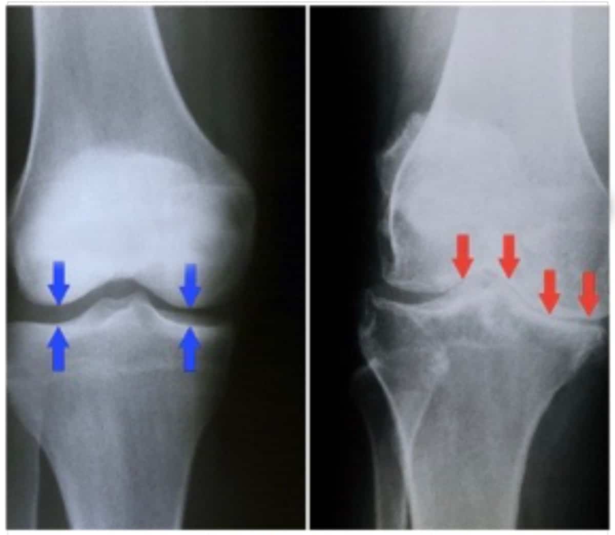
Arthritis is typically diagnosed on x-rays. Osteoarthritis is the most common form of arthritis and is related to wear-and-tear processes, genetics, injuries, and it is a normal part of the aging process. An arthritis joint will demonstrate narrowing of the space between the bones as the cartilage thins, bone spurs on the edges of the joint, small cysts within the bone, and sometimes deformity of the joint, causing it to look crooked. See the x-ray for common findings in osteoarthritis of the hand. The joints closest to the fingertip and the joint at the base of the thumb are the most common joints in the hand affected by osteoarthritis.
X-ray findings in OA of hand:
- joint space narrowing
Recommended Reading: Serum Negative Inflammatory Arthritis
How Does An Mri Work
MRI creates a powerful magnetic field and radio waves to manipulate the position of hydrogen protons within your body. As the protons change position, they give off signals that can be picked up by the MRI scanner. These signals can be used by a computer to make an image of any tissue that is being scanned. Different tissues in the body contain various amounts of water and therefore more hydrogen. As a result, MRI images allow you to see the differences in these tissues.4
Recommended Reading: Can You Get A Rash With Rheumatoid Arthritis
Recommended Reading: What Is The Best Walking Shoe For Bad Knees
Be Cautious With Natural Remedies
Some people pursue complementary therapies such as acupuncture or tai chi when theyre diagnosed with a chronic condition.
If youre seeking a nontraditional avenue, dont keep it a secret from your rheumatologist, and keep in mind that no treatment will be as effective as what your doctor prescribes.
Be especially wary of herbal remedies. There are some alternative therapies that could be dangerous and worsen your symptoms, Dr. Batt says. Its important to tell your doctor everything that youre doing. Personally, I dont have a problem with alternative therapies as long as theyre done in tandem with traditional treatment, at the same time.
What Does Ra Look Like On An Xray
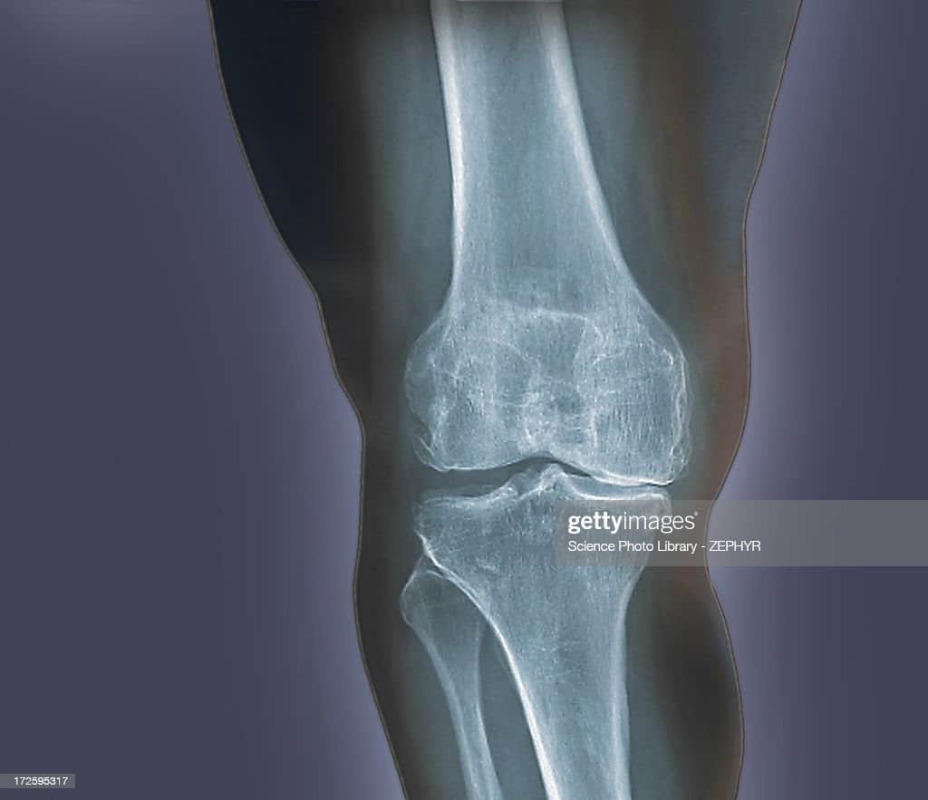
Usually, the earliest sign of RA is a periarticular soft-tissue swelling with a fusiform appearance. Normal fat planes may be obliterated, which occurs as a result of joint effusion, edema, and tenosynovitis. Juxta-articular osteopenia is another early sign, particularly during the acute inflammatory stage.
Read Also: Knee Cartilage Replacement Surgery Recovery Time
What Do Imaging Tests Look For
RA imaging tests look for signs of bone erosion, inflammation and swelling, tissue damage and overall joint deterioration. They are used for the evaluation of RA symptoms and in monitoring the progression of the disease over time.
Imaging tests can show where the patterns of deterioration are beginning to form around the joints. Bone erosion is a very serious concern in RA patients and can be irreversible. Imaging tests identify this early on so that targeted treatment can begin and attempt to prevent further damage to the joint.
If imaging tests do not find any specific or conclusive signs of bone deterioration, it doesnt entirely rule out a RA diagnosis. It could mean instead that the disease is in its early stages and hasnt yet produced any permanent bone damage.
Imaging tests ultimately provide an opportunity to treat RA symptoms as soon as possible. Treatment is far more effective in reducing pain, stiffness, swelling and bone erosion when started in the early stages of the disease.
Bone Marrow Edema And Erosions

Based on a number of prior reports, subchondral and often peripheral marrow edema depicted on MR images in RA patients is a strong predictor of future erosive disease.24,25 Further, histological studies of bone marrow edema indicate that the altered marrow signal is composed of inflammatory cellular infiltrate and a sterile osteitis, which may predispose to a molecular reaction that activates bone and cartilage cleaving enzymes.26 Although roughly half of the patients with marrow edema go on to develop frank erosions within one year, the altered marrow signal can be reversible.16 Given the histologic background of marrow edema, it is not surprising that marrow edema in RA can enhance after the administration of gadolinium.
On both conventional radiographs and MR imaging, erosions are identified as a focal area of cortical loss, typically with acute margins. The bare areas of the joint are commonly affected, as these areas do not possess a protective layer of cartilage. On MR images, erosions are best depicted on non-fat suppressed short TE sequences. Recent erosions can be identified when there is enhancing synovial tissue within the erosion itself, as seen in the ulnar styloid in the introductory case. The detection of bone erosions in early RA correlates with a poor long-term outcome for the patient.16
Figure 9:
Also Check: How Do You Relieve Knee Pain
How Does A Normal Joint Work
A joint is where two bones meet. Most of our joints are designed to allow the bones to move in certain directions and within certain limits.
For example, the knee is the largest joint in the body and one of the most complicated. It must be strong enough to take our weight and must lock into position, so we can stand upright.
It also has to act as a hinge, so we can walk, and needs to twist and turn when we run or play sports.
The end of each bone is covered with cartilage that has a very smooth, slippery surface. The cartilage allows the ends of the bones to move against each other, almost without rubbing.
The joint is held in place by the synovium, which contains thick fluid to protect the bones and joint.
The synovium has a tough outer layer that holds the joint in place and stops the bones moving too far.
Strong cords called tendons anchor the muscles to the bones.