Articular Cartilage And Synovial Fluid
Articular cartilage, also known as hyaline cartilage, is a type of slick, hard, bone-like, flexible connective tissue that covers the surface ends of the tibia and femur at your knee joint, reducing friction and allowing the bones to move easily against one another. It is generally 1/8 to 1/4 inch thick.
Synovial fluid is a thick, stringy, yolk-like fluid that is secreted by the synovial tissue inside the knee capsule. It nourishes the cartilage and lubricates the knee joint.
Wear and tear in the knee joint can cause the protective cartilage to begin to break down. When this occurs it is called osteoarthritis and if left untreated, it can lead to permanent cartilage loss and chronic knee pain.
Our customer service lines are open 5 days a week helping people understand their injuries and how to treat them. Simply call toll free to talk with one of our knowledgeable Product Advisors. They have the ability to answer questions and even put together a treatment plan for you.
Product Advisors are available 9:00 am to 5:00 pm Eastern Standard Time Monday to Friday.
How Are Cruciate Ligaments Injured
The anterior cruciate ligament is one of the most common ligaments to be injured. The ACL is often stretched and/or torn during a sudden twisting motion . Skiing, basketball, and football are sports that have a higher risk of ACL injuries.
The posterior cruciate ligament is also a common ligament to become injured in the knee. However, the PCL injury usually occurs with sudden, direct impact, such as in a car accident or during a football tackle.
Anatomy Of The Knee Bones And Joints Of The Knee Anatomy
The anatomy of the knee consists of 3 main bones:
- The femur .
Lateral Knee Anatomy
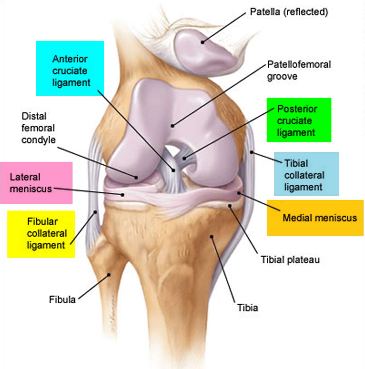
The femur and the tibia are the main movers of the joint to allow for the hinge motion. This connection of the femur and tibia is a joint called the tibiofemoral joint. The patella sits on top of the tibiofemoral joint in a groove in the front of the femur. The patella is a floating bone that works as a fulcrum for the quadriceps muscle to function properly. This joint is called the patellofemoral joint and allows the patella to move up and down, and the knee bends and straightens.
Read Also: How Long To Recover From Knee Surgery
Bones Around The Knee
There are three important bones that come together at the knee joint:
A fourth bone, the fibula, is located just next to the tibia and knee joint, and can play an important role in some knee conditions.
The tibia, femur, and patella, all are covered with a smooth layer of cartilage where they contact each other at the knee joint. There is also a small bone called a fabella, that is often located behind the knee joint.
A type of bone called a sesamoid bone , the fabella is of little consequence to the function of the knee joint. It is only found in about 25% of the population.
What Do The Knee Ligaments Do

Knee ligaments have several important jobs. They:
- Absorb shock when the foot strikes a surface.
- Connect the thigh bone to the lower leg bones.
- Keep the bones in the proper position.
- Prevent the knee from twisting or collapsing.
- Stabilize the knee joint.
- Stop the knee from moving in any unsafe or unnatural directions.
Read Also: Heat Or Ice For Arthritic Knee
Common Knee Ligament Injuries
Anything that stretches the knee ligaments beyond their elastic limit will cause the ligament to tear. Ligament tears vary from a minor sprain where only a few fibers are torn to major tears where the ligament ruptures completely.
You can find out more about knee ligament injuries, including the causes, symptoms, treatment and injury prevention:
How Do The Anatomy Of Knee And Lower Leg Affect Movement
The knee is a hinge joint that sits between the thigh and the shin. It functions the same as a hinge on a door and sometimes gets a creaky as a hinge can. This joint allows the legs to bend and straighten, necessary for walking, going up and downstairs, going from sitting to standing, running, and jumping. The knee’s anatomy consists of many structures from the bones, tendons, and ligaments to the cartilage and muscles to help the knee function.
If you want to learn more about knee anatomy, please watch this knee anatomy video or this article Knee JOINT Anatomy.
Also Check: What Causes My Knee To Suddenly Give Way
Ligaments In The Knee
Ligaments are strong, tough bands that are not particularly flexible. The function of ligaments is to attach bones to bones and to help keep them stable. In the knee, they give stability and strength to the knee joint as the bones and cartilage of the knee have very little stability on their own.
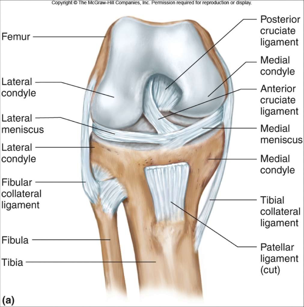
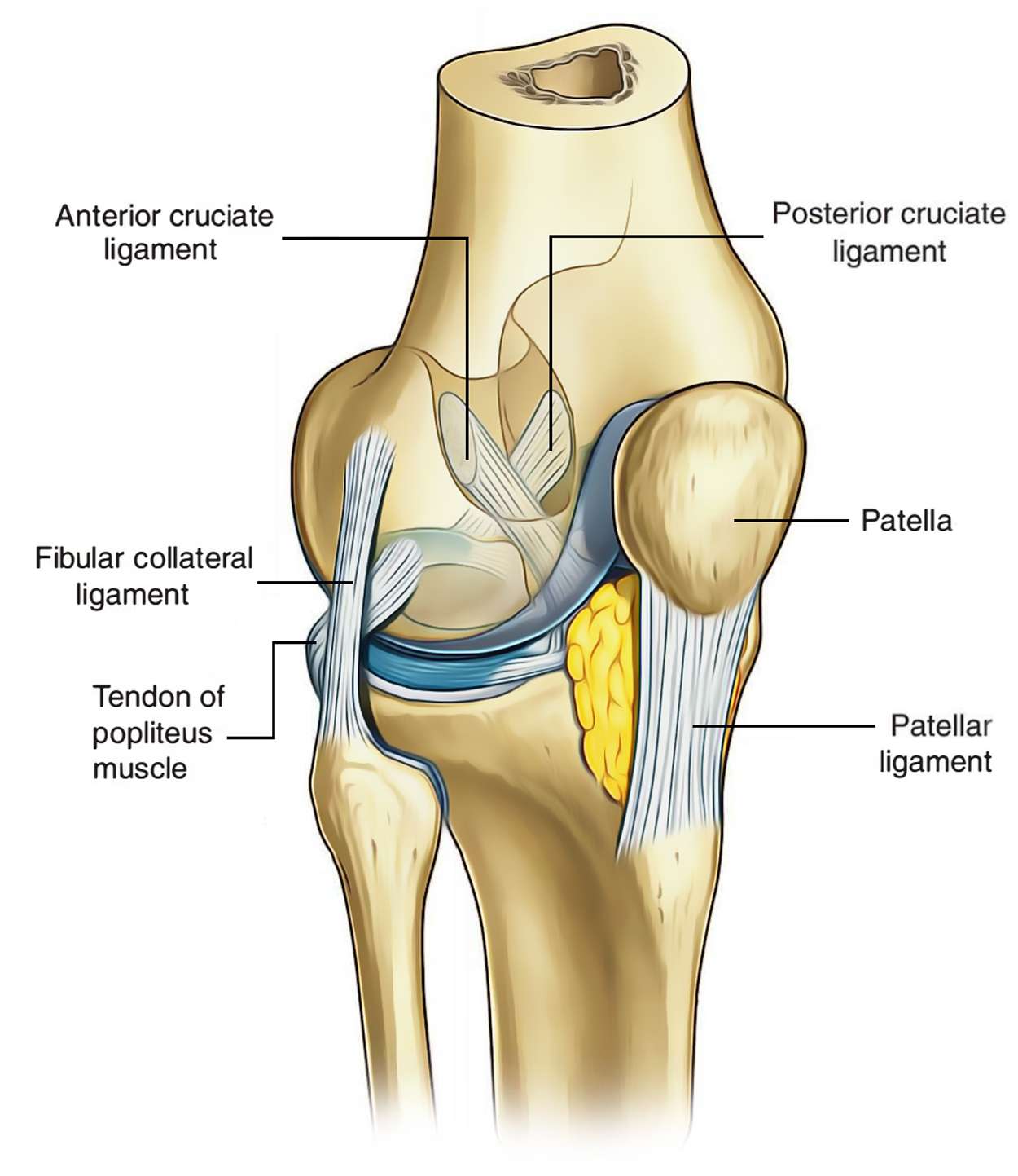
The pair of collateral ligaments keeps the knee from moving too far side-to-side. The cruciate ligaments crisscross each other in the center of the knee. They allow the tibia to swing back and forth under the femur without the tibia sliding too far forward or backward under the femur. Working together, the 4 ligaments are the most important in structures in controlling stability of the knee.
Osteokinematics And Range Of Motion
The ligaments and menisci provide static stability and the muscles and tendons dynamic stability.
The main movement of the knee is flexion – extension. For that matter, knee act as a hinge joint, whereby the articular surfaces of the femur roll and glide over the tibial surface. During flexion and extension, tibia and patella act as one structure in relation to the femur. The quadriceps muscle group is made up of four different individual muscles. They join together forming one single tendon which inserts into the anterior tibial tuberosity. embedded in the tendon is the patella, a triangular sesamoid bone and its function is to increase the efficiency of the quadriceps contractions. Contraction of the quadriceps pulls the patella upwards and extends the knee.Range of motion: extension 0o. The hamstring muscle group consists of the biceps femoris, semitendinosus and semimembranosus. They are situated at the back of the thigh and their function is flexing or bending the knee as well as providing stability on either side of the joint line.Range of motion: flexion 140o.
Secondary movement is internal – external rotation of the tibia in relation to the femur, but it is possible only when the knee is flexed.
Don’t Miss: How Do I Fix Knee Pain
When Should I See A Healthcare Provider For A Sprained Knee
Damage to a knee ligament can weaken the knee joint, increasing the chances that youll injure yourself again.
Talk to a healthcare provider if you have:
- Looseness or weakness in the knee.
- Loss of feeling in the knee or leg.
- Pain on the inside or outside of the knee.
- A popping or snapping noise.
- Repeat knee injuries.
- Swelling around the knee joint.
- Trouble putting weight on that leg.
A note from Cleveland Clinic
Knee ligaments are bands of tissue that connect the thigh bone in the upper leg to the lower leg bones. There are four major ligaments in the knee: ACL, PCL, MCL and LCL. Injuries to the knee ligaments are common, especially in athletes. A sprained knee can range from mild to severe. Talk to a healthcare provider if you have a severe knee injury or repeat injuries. Proper diagnosis and treatment can help prevent pain and future injuries.
Orthopedics/traumatology Tiefenau Hospital Of The City Of Bern Bern Switzerland
H.-U. Stäubli
-
Book Title: The Knee and the Cruciate Ligaments
-
Book Subtitle: Anatomy Biomechanics Clinical Aspects Reconstruction Complications Rehabilitation
-
Editors: R. P. Jakob, H.-U. Stäubli
-
DOI: https://doi.org/10.1007/978-3-642-84463-8
-
Copyright Information: Springer-Verlag Berlin Heidelberg 1990
-
Softcover ISBN: 978-3-642-84465-2
-
Number of Pages: XXIV, 637
-
Number of Illustrations: 247 b/w illustrations
Also Check: What Doctor Do You See For Knee Problems
Cartilage Of The Knee
There are two types of the cartilage of the knee joint:
Ligaments & Joint Capsule

The joint capsule has thick and fibrous layer superficially and thinner layers deeper. This along side the capsule ligaments enhances she stability of the knee. As with all of the structures that from the knee they are under most tension therefore more stable in an extended position in comparison to the laxity present in a flexed position . Inside this capsule is a specialized membrane known as the synovial membrane which provides nourishment to all the surrounding structures. The synovial membrane produces synovial fluid which lubricates the knee joint. Other structures include the infrapatellar fat pad and bursa which function as cushions to exterior forces on the knee. The synovial fluid which lubricates the knee joint is pushed anteriorly when the knee is in extension, posteriorly when the knee is flexed and in the semi flexed knee the fluid is under the least tension therefor being the most comfortable position if there is a joint effusion.
The ligaments of the knee maintain the stability of the knee. Each ligament has a particular function in helping to maintain optimal knee stability.
|
|
|
Superior gluteal |
|
You May Like: Will Losing Weight Help My Knee Pain
Muscles And Tendons Of The Knee
Many muscles affect the knee, but the main muscles that allow for the knee to perform its main functions are:
- Quadriceps: A group of 4 muscles that sits on the front of the thigh. These muscles are responsible for allowing the knee to straighten. This movement is necessary for standing from a seated position, bringing your leg forward when walking, and kicking a ball! The two patellar tendons attach the quad to the patella. These tendons can also rupture during sports.
Quadriceps Muscle diagram
What Are The Ligaments Of The Knee
There are two main types of ligaments in your knee:
- Collateral ligaments: The two collateral ligaments are like straps on each side of your knee. The medial collateral ligament is on the inner side of your knee. It attaches the thigh bone to the shin bone . The lateral collateral ligament is on the outer side of your knee. It connects your femur to your calf bone . The collateral ligaments prevent the knee from moving side to side too much.
- Cruciate ligaments: The two cruciate ligaments are inside your knee joint and connect your femur to your tibia. They cross each other to create an X. The anterior cruciate ligament is located toward the front of the knee. The posterior cruciate ligament is behind the ACL. The cruciate ligaments control the way your knee moves front to back.
Read Also: What’s Good For Your Knees
How Is A Knee Ligament Injury Diagnosed
In addition to a complete medical history and physical examination, diagnostic procedures for a knee ligament injury may include the following:
-
X-ray. A diagnostic test that uses invisible electromagnetic energy beams to produce images of internal tissues, bones, and organs onto film to rule out an injury to bone instead of, or in addition to, a ligament injury.
-
Magnetic resonance imaging . A diagnostic procedure that uses a combination of large magnets, radiofrequencies, and a computer to produce detailed images of organs and structures within the body can often determine damage or disease in bones and a surrounding ligament or muscle.
-
Arthroscopy. A minimally-invasive diagnostic and treatment procedure used for conditions of a joint. This procedure uses a small, lighted, optic tube that is inserted into the joint through a small incision in the joint. Images of the inside of the joint are projected onto a screen used to evaluate any degenerative and/or arthritic changes in the joint to detect bone diseases and tumors to determine the cause of bone pain and inflammation.
Cartilage Of The Knee Joint
There are two main types of cartilage in knee anatomy: articular cartilage and the meniscus.
- Articular cartilage covers the bones’ ends and allows for the bones to slide and glide on each other without friction. This is the stuff you need to keep from getting the creaking and cracking of the joints. When this starts to wear down, arthritis will set in. Sometimes this cartilage is damaged with an ACL tear. The amount of trauma from the ACL injury can lesions to the cartilage of the joint or bones of the knee. This can be addressed during the surgical procedure.
Image of articular cartilage and meniscus
- Meniscus: 2 thick pieces of cartilage that sit on the tibia between the femur and tibia. These are C-shaped that allow for improved congruence of the joint. Tears in these structures can cause pain, swelling, and sometimes catching and locking the knee joint. During surgery, the meniscus can be repaired or debrided. This is usually determined by the age of the patient, where the tear occurred and the amount of damage to the meniscus. To learn more, Read this Article about Meniscus Injuries.
Don’t Miss: Blood Clot Behind Knee Treatment
Muscles Around The Knee
The muscles around the knee help to keep the knee stable, well aligned, and moving. There are two main muscle groups around the knee: the quadriceps and the hamstrings. The quadriceps are a collection of 4 muscles on the front of the thigh and are responsible for straightening the knee by bringing a bent knee to a straightened position. The hamstrings are a group of 3 muscles on the back of the thigh that provide the opposite motion by bending the knee from a straightened position.
The iliotibial band is a broad tendinous extension of the tensor fascia lata and gluteus maximus that also helps to stabilize the knee.
What Are The Treatment Options For Knee Injuries
Ice
Placing a bag of ice directly on the knee for 20 minutes prevents swelling and inflammation from occurring inside the knee.
Anti-inflammatory medicines
Over-the-counter medications such as Aleve, Advil, Motrin, and aspirin can help reduce swelling and pain.
Injections
In the specialists office, medication is injected directly into the joint. Injections can provide lasting relief of pain and swelling.
Physical therapy
A therapist can work with you to first control your pain and inflammation. They then will help you regain your strength and range of motion.
Wellness
Start with a free wellness consultation to explore your goals and what tools are available to you.
Integrated physical therapy and wellness
Our team of physical therapists, performance specialists, and registered dietitians works collaboratively with you to design an integrated rehabilitation plan that will help you reach your full potential.
Surgery
If you fail to improve after nonsurgical care, your specialist may wish to intervene surgically. Your specialist can discuss the details of the surgery with you should it become necessary.
Read Also: What Is The Best Knee Brace To Buy
Ligaments And Tendons Of The Knee
The knee has 4 main ligaments:
- Medial collateral ligament : On the inside of the knee closer to the midline.
- Lateral collateral ligament : Is on the outside of the knee.
- An anterior cruciate ligament : Inside of the knee and crosses to the front.
- A posterior cruciate ligament : Inside of the knee and crosses to the back.
The MCL and the LCL sit on the sides of the knee, and they help give stability to the knee if your knee gets hit from the sides. Knee bones, ligaments, and meniscus
The ACL and PCL are inside the knee and cross each other as they run front to back and vise versa. These 2 ligaments are responsible for giving the knee stability from front to back.
An ACL injury is probably one of the most recognized injuries in sports and, most of the time, requires a surgical repair that has a long recovery time. A full recovery after an ACL reconstruction is usually between 6 to 9 months depending on the patient and the other structures injured.
The unhappy triad is referred to when the ACL, MCL and Medial Meniscus are all injured at the same time. .
Tendons are where muscles attach to the bones of the knee. There are numerous tendons in the knee. The tendons which are prone to injuries of the knee are the Patellar Tendon and the Quadriceps Tendon. These patellar tendons can rupture or tear and they can also get tendonitis.
How Are Knee Sprains And Tears Classified

A healthcare provider will grade your injury by how severe it is and what symptoms you have:
- Grade 1: A grade 1 injury to a knee ligament is a minor sprain. The ligament is overstretched or just slightly torn. With a grade 1 knee strain, youll have minimal pain, swelling or bruising. Youll still be able to put weight on the affected leg and bend the knee.
- Grade 2: A grade 2 knee sprain is a moderate tear of the ligament. Signs include bruising, swelling and some pain. With a grade 2 injury, youll have some difficulty putting weight on the leg or bending the knee.
- Grade 3: A grade 3 injury is a complete tear or rupture of the knee ligament. Grade 3 injuries often involve more than one knee ligament. With this level of injury, you will experience severe bruising, swelling and pain. You wont be able to put weight on the leg or bend the knee.
Also Check: Why Does My Knee Hurt When It’s Cold