How Imaging Tests Help Diagnose Ra
None of the imaging tests on their own can reliably diagnose RA. RA is a clinical diagnosis, meaning that results from imaging tests is used by your doctor in combination with the assessment of physical symptoms, blood tests, and medical history to diagnose RA.
Imaging tests are helpful tools for diagnosis and providing a clear medical picture of your present condition. Imaging tests are also used post-diagnosis to continue to monitor your level of bone erosion. Imaging tests can indicate the severity and speed of the diseases progression.
Also Check: Why Is My Arthritis Acting Up
Complications Of Rheumatoid Arthritis
There are many complications of RA, which occur predominantly in the setting of longstanding disease.
Stenosing tenosynovitis is a frequent complication of longstanding RA this condition results in impaired tendon movement in the setting of chronic inflammation of the tendon sheath. One study found stenosing tenosynovitis to occur in one third of RA patients with an average disease duration of 5.9 years.39 Stenosing tenosynovitis is most commonly seen in the hand and wrist, particularly involving the tendons in the extensor compartments. With MR imaging, stenosing tenosynovitis manifests as bead-like dilatation of the tendon sheath with areas of synovitis.
Rotator cuff tears are a common complication of RA. Massive rotator cuff tears, characterized by full-thickness and full-width tears of two or more rotator cuff tendons, are best diagnosed with MR imaging .
Figure 16:
A coronal fat-suppressed T2-weighted image demonstrates a full-thickness and full-width tear of the supraspinatus tendon with retraction of torn fibers to near the glenohumeral joint . A fat-suppressed proton density-weighted axial image reveals a full thickness tear of the subscapularis tendon. Rice bodies are apparent in the subdeltoid bursa. A sagittal T1-weighted image demonstrates marked atrophy of the supraspinatus muscle and mild atrophy of the infraspinatus muscle .
Figure 17:Figure 18:
Figure 21. Classic sites of involvement in the hand and wrist in RA .
Youre Trying To Cope With Knee Osteoarthritis By Yourself
People with knee osteoarthritis often know that healthy lifestyle habits like exercise and weight control are recommended, but they arent implementing them, Dr. Garver says. His research, which was published in the October 2014 issue of The Journal of Rheumatology found that meeting with others who have osteoarthritis and sharing similar challenges can help motivate people to change their habits and add an exercise routine into their life.
Recommended Reading: What Is The Mcl In The Knee
You May Like: Why Does The Back Side Of My Knee Hurt
Differences Between Ra And Oa
As rheumatoid arthritis progresses, the more severe symptoms appear. Following are some differences between rheumatoid arthritis and osteoarthritis.
- RA is an autoimmune disease so the immune system is compromised, but in osteoarthritis autoimmune issues are not present.
- RA symptoms have a rapid onset, while OA progresses slowly.
- RA affects joints throughout the body, while OA affects mostly knees, small finger joints, thumb and hips .
- RA creates systemic symptoms, like fatigue and low-grade fever, while OA is localized to a joint.
- RA is symmetrical, so both sides of the body are affected in similar joints, while OA affects individual joints.
- RA morning stiffness may last longer than 30 minutes, while the stiffness people with OA experiences is likely to ease within 30 minutes.
Special Forms Of Rheumatoid Arthritis

Adult Still Syndrome
This polyarticular, systemic course of chronic polyarthritis is accompanied by swelling of the liver, spleen, and lymph nodes, high fever, leukocytosis, iridocyclitis, and polyserositis , as well as polyarthralgia and myalgia. Rheumatoid factors are negative. Articular symptoms are often first seen at later stages of the disease, usually as symmetric polyarthritis.
Felty Syndrome
This includes variants of seropositive rheumatoid arthritis with strong activation of the lymphoreticular system, splenomegaly, and lymphadenopathy. Laboratory findings show leukopenia or granulocytopenia and thrombopenia.
Sjögren Syndrome
This autoimmune adult exocrinopathy occurs predominantly amongwomen in menopause. There is a decrease in tear and saliva secretion . Focal lymphocytic or plasma-cell infiltration can cause internal organs, particularly the large salivary glands, to swell. This combination of symptoms without articular involvement is referred to as sicca syndrome. The primary form, which can progress to B-cell lymphoma, is differentiated from the secondary form, which involves a combination of the sicca syndrome and rheumatoid arthritis or collagenosis.
Caplan Syndrome
This is a combination of pneumoconiosis with rheumatoid arthritis. Both the pneumoconiosis and the synovial disease apparently reflect a rheumatoid reaction of the mesodermal tissue.
Juvenile or Special Forms of Rheumatic Arthritis
Diagnostic Imaging
Radiography
2a-cabc3a, bab4a, b54
Recommended Reading: How To Diagnose Knee Problems
Utility And Technical Considerations Of Mr Imaging In Ra
The utility of magnetic resonance imaging in the evaluation of RA has been well described in the literature.15 MR imaging clearly offers increased sensitivity to soft tissue and marrow abnormalities and provides the clinician valuable diagnostic information that may significantly alter the management of RA, particularly in the initial stages, allowing earlier treatment and significant reduction in the morbidity of the disease.16 Reported data have indicated that approximately one third of patients in clinical remission with non-tender or non-swollen joints have evidence of synovitis on MR images.17 The sensitivity of MR imaging when clinical findings are absent or subtle is important as conventional radiographs are unrevealing at such a time. Furthermore, subclinical synovitis is the predominant finding and may progress in severity, leading to bone and cartilage destruction in roughly 47% of patients.18
An Efficient Cnn For Hand X
Bhupesh Kumar Singh
1Department of Electronics and Telecommunication, JSPMs Rajarshi Shahu College of Engineering, Pune 411033, India
2Department of Electronics, Maulana Mukhtar Ahmad Nadvi Technical Campus, Malegaon 423203, India
3Arba Minch Institute of Technology, Arba Minch University, Arba Minch, Ethiopia
Abstract
Hand Radiography is one of the prime tests for checking the progress of rheumatoid joint inflammation in human bone joints. Recognizing the specific phase of RA is a difficult assignment, as human abilities regularly curb the techniques for it. Convolutional neural network is the center for hand recognition for recognizing complex examples. The human cerebrum capacities work in a high-level way, so CNN has been planned depending on organic neural-related organizations in humans for imitating its unpredictable capacities. This article accordingly presents the convolutional neural network which has the ability to naturally gain proficiency with the qualities and anticipate the class of hand radiographs from an expansive informational collection. The reproduction of the CNN halfway layers, which depict the elements of the organization, is likewise appeared. For arrangement of the model, a dataset of 290 radiography images is utilized. The result indicates that hand X-rays are rated with an accuracy of 94.46% by the proposed methodology. Our experiments show that the network sensitivity is observed to be 0.95 and the specificity is observed to be 0.82.
Recommended Reading: When To Do Knee Replacement Surgery
Rheumatoid Arthritis Vs Osteoarthritis: What Is Common
Rheumatoid arthritis and osteoarthritis have some things in common. The most obvious commonality is that both affect joints. There is no doubt that both can significantly harm the quality of life, if the disease is not purposefully managed. The most significant difference between the two types of arthritis is the underlying cause.
The most familiar type of arthritis is osteoarthritis OA, also called the wear-and-tear disease. Arthritis a degenerative disease develops when the cushioning cartilage in the joint wears out or disintegrates. Pain occurs when bones rub against each other. Sometimes, bone spurs develop, increasing pain and causing inflammation in surrounding tissues.
Another common occurrence is when bits of bone or cartilage float around in the joint, causing painful inflammation. Osteoarthritis will continue to get worse over time, which is why it is called degenerative. Eventually, all the cartilage wears away.
Correlation Of Mri To Histological Findings For The Menisci
The degree of meniscus destruction was classified as grade 4 in the majority of patients, for both the medial and lateral meniscus, with an average degree of meniscal destruction over all specimens of 3.85 the destruction grade of the medial meniscus was 4 and of the lateral meniscus was 3.69. Atrophy and severe destructions of the menisci are, therefore, characteristic features of RA on MRI. The degree of destruction was more severe for the medial meniscus than for the lateral meniscus . Among all specimens, 10 menisci were available , with complete destruction of the menisci in the other specimens. The histological findings of the menisci in RA included : synovial cells on the meniscal surface , fibrosis , fibrinoid necrosis , vascular hyperplasia , lymphocyte infiltration , plasmacyte infiltration , and engulfing and encapsulating calcified debris . Fibrosis, engulfing and encapsulating calcified debris, and fibrinoid necrosis were the most severe histological findings. In cases in which the menisci had been completely destroyed, synovium was identified on the surface of the menisci, which may by the main cause of meniscal destruction.
Don’t Miss: Runner’s Knee Physical Therapy
Turmeric/curcumin Or Curcuma Longa
A popular nutritional supplement well known for its anti-inflammatory effects, turmeric and more specifically its active constituent curcumin is an effective natural treatment for pain but also inflammatory conditions such as rheumatoid arthritis.
Clinical studies indicated that the use of curcumin provided a decrease in pain and stiffness among patients suffering from arthritis and rheumatoid arthritis .
Curcumin has been shown to be a safe and effective treatment for not only joint pain management but it also exerts protective effects in the prevention of joint inflammation .
Turmeric is a medicinal herb and spice that can be used in cooking, though supplementation is likely needed for therapeutic effects and more severe pain management.
Arushi Jain
Arushi Jain, Director , Akums Drugs & Pharmaceuticals Ltd.
What can help in rheumatoid arthritis?
How can vitamin D help in the effective management of rheumatoid arthritis?
Where can we get vitamin D from?
Imaging Tests For Rheumatoid Arthritis
Learn what types of imaging scans are most effective in detecting and monitoring RA.
For decades, X-rays were used to help detect rheumatoid arthritis and monitor for worsening bone damage. In the early stages of RA, however, X-rays may appear normal although the disease is active, making the films useful as a baseline but not much help in getting a timely diagnosis and treatment.
Newer imaging techniques like musculoskeletal ultrasound and magnetic resonance imaging have changed the picture. Both can pick up inflammation and bone erosion not visible on X-rays. This is especially important because early diagnosis and treatment can help forestall future joint damage.
Imaging in Diagnosis
RA is diagnosed based on a physical exam, medical history and certain lab results. Imaging tests arent diagnostic themselves but can support other findings, explains rheumatologist Flavia Soares Machado, MD, of the Universidade Federal de São Paulo school of medicine in Brazil.
You can see the same bone erosion and synovial lining changes in other rheumatic diseases, such as lupus and psoriatic arthritis . So the clinical history and physical examination are still important, with careful evaluation of the pattern of joint involvement and some blood tests to make the diagnosis, she says.
Predicting Outcomes
Recommended Reading: Are There Restrictions For Returning To Work After Knee Replacement
Articles On Natural Remedies For Ra Pain
Medication and other forms of treatment play a major role in managing rheumatoid arthritis. Supplements arent a treatment they dont promise to cure or treat any condition, and they dont have to meet the same standards as medications. But if youre looking into trying some in addition to your treatment plan, heres what you should know.
First, be sure you talk to your doctor before you begin using any supplement or herbal medication. They may interact with other drugs, supplements, or herbal medicines you are taking and cause serious side effects. They may also put you at a higher risk for certain conditions.
While Both Types Of Arthritis There Are Notable Differences

Osteoarthritis is the most common type of arthritis. Rheumatoid arthritis is recognized as the most disabling type of arthritis. While they both fall under the arthritis umbrella and share certain similarities, these diseases have significant differences.
- 14. Musculoskeletal
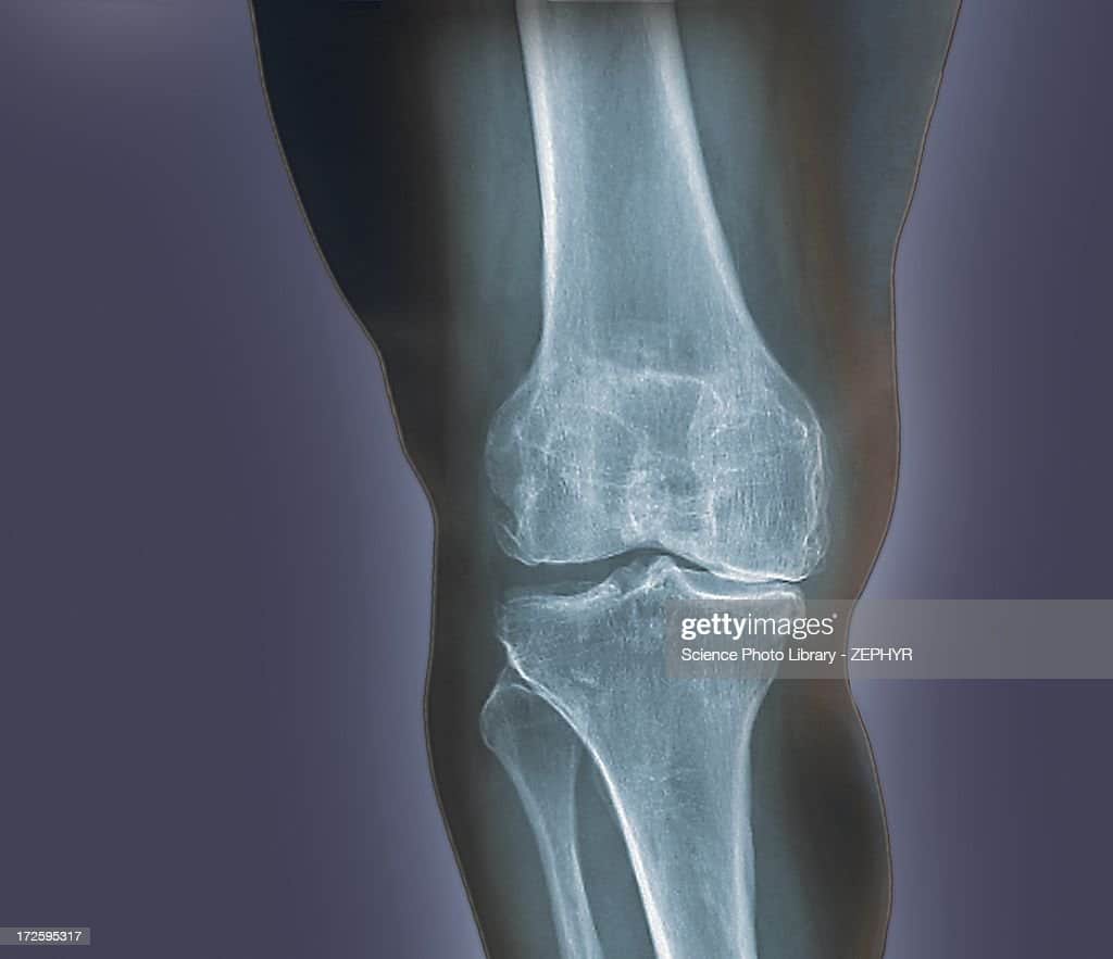
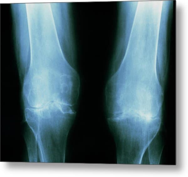
Arthritis on X-Ray is clearly seen on the image, and the doctors often use the result from X-Ray to confirm it. The X-Ray helps in obtaining the images of tissues, organs, and other body structures.
The most common form of arthritis, osteoarthritis, is associated with injuries, wear-and-tear processes, and genetics. An arthritis joint will demonstrate the narrow bone spaces due to various reasons.
Read them carefully to arrive at the decision of buying the best product on the market. The pregnant women must inform the technician about their pregnancy because the exposure to radiation may cause harm to the fetus, so it must be minimized.
There could be a requirement to remove some clothes, depending on the body parts to be tested. The energy produced by the machine will pass through the body part that is being X-rayed.
The bones along with various other dense areas will be showed up as lighter shades of gray to white. These areas will appear as dark gray to black color.
The study shows that arthritis is forcing people to retire early from their workplace. The treatment methods suggested by orthopedist have helped out many patients suffering from osteoarthritis from the past until now.
Don’t Miss: What Can Make Your Knee Hurt
Arthritis In Neck And Shoulders
Arthritis of the neck, also called cervical spondylosis, affects more than 85% of people over the age of 60. Pain and stiffness in the neck are the most common symptoms. They often respond well to conservative treatment like pain medications and physical therapy.
Symptoms of neck arthritis can worsen with looking up or down for a sustained duration or with activities like driving and reading that involve holding the neck in the same position for a prolonged period of time. Rest or lying down often help to relieve symptoms.
Other symptoms of neck arthritis include:
Arthritis of the shoulder can develop over time from repetitive wear-and-tear or following a traumatic injury such as a shoulder fracture, dislocation, or rotator cuff tear. The most common symptoms of shoulder arthritis include pain, stiffness, and loss of range of motion. As arthritis progresses, any movement of the shoulder can cause pain.
If symptoms do not improve with conservative measures, surgical methods may be used to manage symptoms of shoulder arthritis. Surgical options include:
rustycloud / Getty Images
Other Tests For Seropositive Rheumatoid Arthritis
Blood tests are not only used to detect RF and anti-CCP antibodies. Theyre also used to reveal if you have:
- Anemia, or low red blood cell count, which occurs in up to half of people with RA
- A high erythrocyte sedimentation rate, also known as a sed or ESR rate, a crude measure of inflammation in your body
- High C-reactive protein levels, another marker of inflammation
Aside from blood tests, an X-ray can help your doctor determine the degree of destruction in your joints, but may only be useful when RA has progressed to a later phase.
You May Like: How To Help Aching Knees
Reducing The Need For Treatment
NSAIDs are often recommended for short-term pain relief for patients with RA. These medicines exert their analgesic effects by inhibiting COX, an enzyme responsible for pain and inflammation.3 As their use is associated with increased risk of upper gastrointestinal and CV events, NSAIDs should be used in the lowest dose possible needed to reduce pain, and for short periods only.10,11 A meta-analysis concluded that n-3 PUFAs at doses > 2.7 g/day for > 3 months significantly reduced NSAID use by patients with RA with no heterogeneity between studies .3
The effects of cod liver oil supplementation on NSAID requirements for patients with RA are also promising.12
Distribution Of Articular Involvement
Radiographs provide a global assessment of regional arthritis involvement. Although the distribution of radiographic abnormalities in RA is somewhat variable, it is generally symmetrical and polyarticular. RA targets synovial joints thus the distribution of involvement will correlate with the locations of synovial joints and the synovial lining in the joint. There is a predilection for symmetric involvement of the distal extremities, hands, wrists, and feet, but the elbows, knees, shoulders, and hips are often affected. In the axial skeleton, imaging abnormalities in the articulations of the cervical spine are more often encountered than are changes in the thoracolumbar spine or sacroiliac joints.
Don’t Miss: Knee Pain On Front Of Knee
Oa Is Diagnosed With X
Both OA and RA require you to give a medical history and undergo a clinical exam for diagnosis. But for diagnosing OA, X-rays are also important, says Dr. Ashany. X-ray images can show if the space between the bones is becoming narrower, a sign of cartilage loss. And they can reveal the presence of those bony growths called osteophytes. Magnetic resonance imaging may also be used to detect more detailed changes in the cartilage and surrounding tissues, says Dr. Askari.
Which Body Parts Are X
RA begins as a peripheral disease that has a strong affinity for the hands. Plus, since there are more joints in the hands than any other region of the body, doctors often order x-ray images of the hands firstthen the feet.
There are more than 30 joints in the hands, so getting x-rays of the hands provides a very good diagnostic yield, says Dr. Magnati. If the x-rays show bone erosions and narrowing of the joints at the base of your fingers near the palm, the knuckles, and the uppermost joint of the thumb , thats classic RA.
RA tends to affect joints in pairs, so doctors typically order x-rays of both hands and both feet . The other area that can be involved in RA is the neck, where the spine connects to the skull, says Dr. Burk.
Also Check: What Is Acl Knee Surgery
Best Vitamins For Stiff Joints
As people start aging, they may experience joint stiffness. Most experience this joint stiffness in the morning when they wake up and it impacts mobility for a short amount of time. However, in some cases, it may be more serious as one experiences inflammation and discomfort, which makes walking or standing painful.
Get a checkup or blood test from the doctor before consuming any supplements to see what your body is lacking. You can then best supplement that said lack with the relevant and recommended type and dosage.
The best vitamin for joints is vitamin D consumption. This vitamin can be found in fish oil or by going out into the sunlight for at least 30 minutes a day.
If vitamin is still lacking, you can try joint pain supplements such as NOW Vitamin D-3.
You may also try estrogen. Estrogen is said to be the best defence against knee osteoarthritis.
Vital for musculoskeletal health, including joint health, estrogen helps postmenopausal women with low levels of estrogen as women at this age may complain about joint pain and stiffness as their primary menopausal symptom.
For women, we recommend taking Wakunaga Estro Logic as it helps maintain normal estrogen levels as well as alleviate symptoms associated with menopause.
Of course, before taking any supplements, you should always consult with your doctor first in case there are any side effects or it is detrimental to your health. Although taking joint vitamins is generally safe, an overdose is never a good idea.