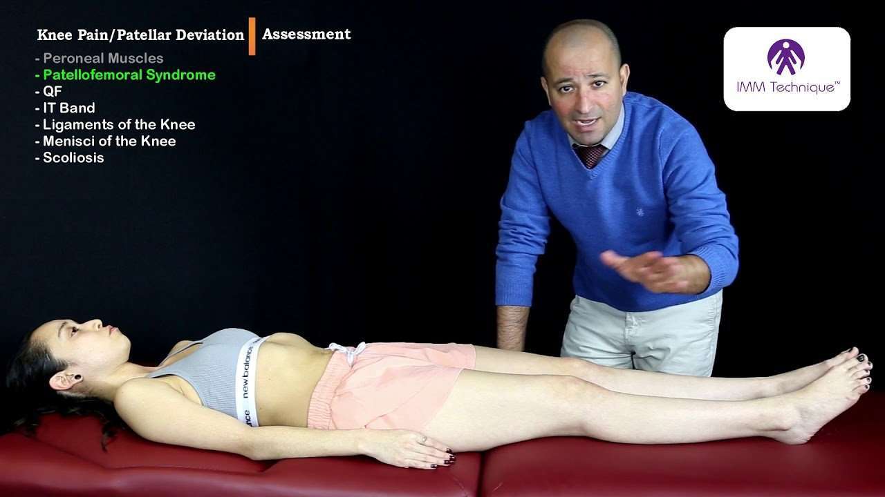
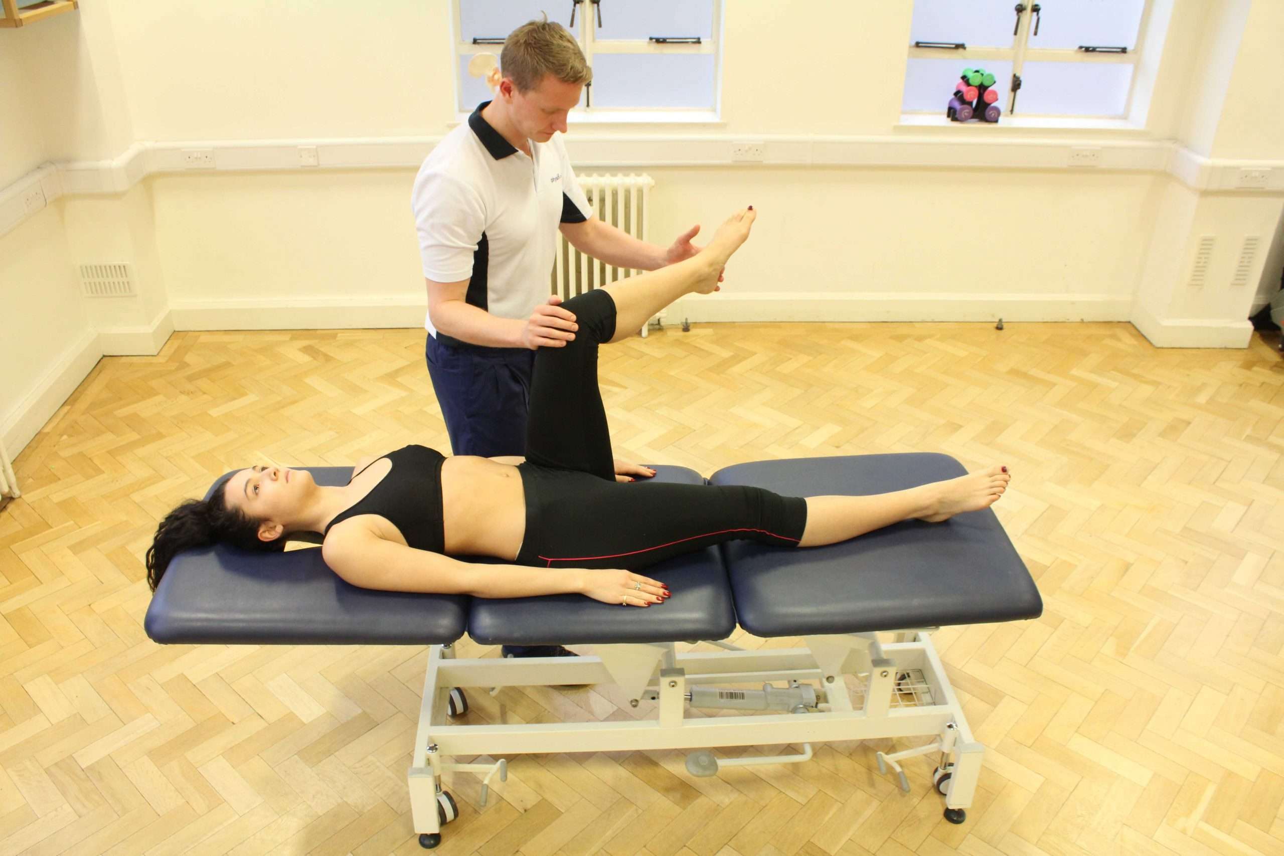
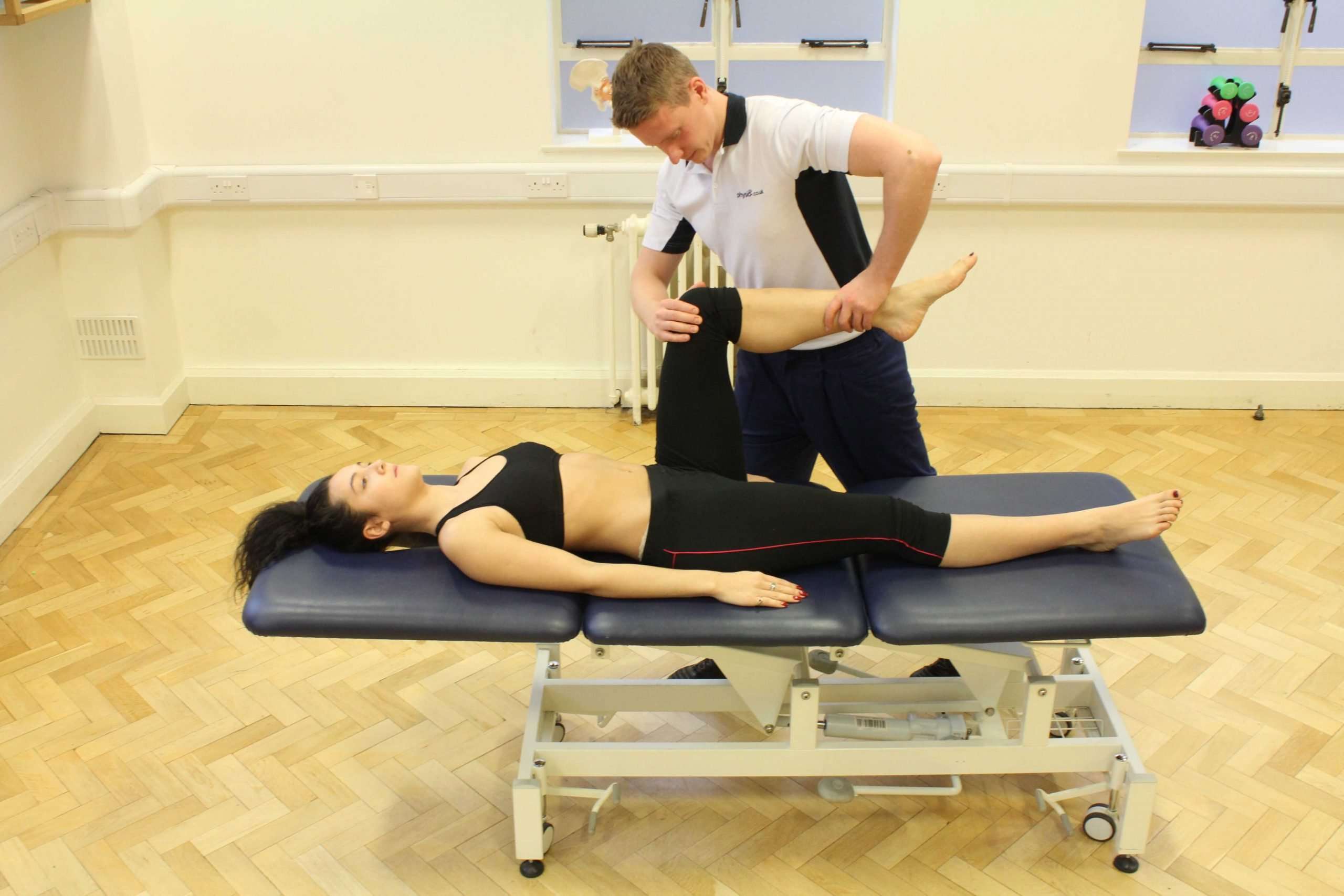
Palpation Of The Extended Knee
With the patients leg straight and relaxed, systematically palpate the joint lines and surrounding structures of each knee joint.
Patella
1. Assess the medial and lateral border of the patella for tenderness by stabilising one side of the patella and palpating the other with a fingertip:
- Tenderness may represent injury or patellofemoral arthritis.
- If the patient appears apprehensive, developing tension in the muscles of the leg as you begin to mobilise the patella , it may suggest a history of recurrent patellar dislocation which the patient is anticipating .
2. Palpate the patellar ligament for tenderness suggestive of tendonitis or rupture.
Medial and lateral joint lines
1. Palpate the medial and lateral joint lines of the knee including the collateral ligaments for evidence of tenderness which may suggest:
- Fracture
- Meniscal injury
- Collateral ligament injury
2. Palpate the quadriceps tendon for tenderness suggestive of tendonitis or rupture.
- Assess and compare knee joint temperature
- Palpate the quadriceps tendon
- Palpate the borders of the patella
- Palpate the patella tendon
Patellar apprehension test
The patellar apprehension test is not usually performed in an OSCE, but its useful to understand how the test is carried out.
With the patients knee fully extended lateral pressure is applied to the patella whilst simultaneously slowly flexing the knee joint. The presence of active resistance from the patient is suggestive of previous patellar instability and dislocation .
Podiatrist At Podiatry First
Here we are about to go through some common orthopaedic tests for knee injuries.
Please keep in mind these are purely for educational purposes. Assessing knee injuries without any kind of training and experience can result in further injury!
The knee is the largest and considered the most complex joint, in the human body. It acts as a hinge that allows your lower leg and foot to swing back and forth while walking. The knee continually accommodates impact as ground reaction forces are put through the joint. Subsequently, the knee is prone to patho-mechanical errors caused by factors such as malalignments, abnormal muscle health, foot type, genetics and even developmental issues.
A complete physical examination of the knee is always done for a knee complaint, whether the complaint is from a recent or sudden injury or from long-lasting or recurrent symptoms .
Due to its intricate nature, to correctly diagnose a knee injury several orthopaedic tests are utilized by health professionals to narrow the pathology or injury down and come up with a diagnosis. Some such tests are as follows.
Assessment Of Quadriceps Femoris Bulk
Reduced bulk of the quadriceps femoris is very suggestive for knee pathology. Knee pain and/or weakness that limits its use will result in wasting. Using a measuring tap, identify a point roughly 20cm superior to the tibial tuberosity on one limb. Record the girth of the quadriceps femoris muscle at this point and then repeat this on the other limb. If the girths are similar, this suggests either no, or bilateral pathology affecting both knees.
Read Also: How Much Does Aflac Pay For Knee Surgery
Measurement Of Quadriceps Bulk
Quadricepswasting is commonly associated with knee joint pathology occurring secondary to disuseatrophy. Wasting will often be apparent on inspection, however subtle wasting may only be detectable by comparativemeasurement of leg circumference.
To measure the circumference of the leg in the region of the quadriceps place a measuring tapearound each leg at a point approximately 20cm above the tibial tuberosity.
Record the circumference of each leg and compare to see if there is a significant difference indicative of quadriceps wasting.
Mechanical Or Heat Sensitivity Of The Paw

Von Frey filaments and a modified Randall-Selitto analgesiometer have been used to assess the mechanical sensitivity of the hindpaw in animals with knee joint arthritis. Typically, paw withdrawal thresholds are measured in response to increasing pressure stimuli applied to the plantar surface by von Frey filaments or to the dorsal surface by a wedge-shaped probe of a Randall-Selitto analgesiometer. Rats with knee joint arthritis induced by MIA have decreased PWT 1) for several weeks on the affected limb measured with either technique, but show little dynamic allodynia assessed by stroking the plantar surface of the paw. Surgically induced knee joint arthritis appears to be more sensitive to von Frey hair testing than to Randall-Selitto analgesiometry . Bilateral decreases of mechanical PWT measured with von Frey filaments occur in the K/C knee joint arthritis model .
These tests assess secondary hyperalgesia or allodynia, which has been reported in patients with osteoarthritis but is not very common . The following direct measures of knee joint pain have been developed recently.
Don’t Miss: How To Get Rid Of Dark Spots On Knees Fast
How To Measure Knee Rom Without A Goniometer
If you dont have a goniometer, you can still assess your knee ROM. This can be really helpful for seeing what progress you are making with your rehab after a knee injury. It wont be as accurate as using a goniometer, but it does give you somewhere to start.
If you are wanting to guestimate the range of movement at your knee, try this:
Estimating Knee Extension ROM
- Lie on your back on a firm surface
- Push your knee down into the floor
- Slide your hand, palm down, underneath your knee. If you can:a. Just get a couple of your fingers underneath with difficulty = 0o extensionb. Just slide all your fingers underneath = +5o degrees i.e. lacking 5o extensionc. Easily slide your whole hand underneath = +10o i.e. lacking 10o extensiond. Cant get any of your fingers underneath = -5o or more i.e. hyperextension
Estimating Knee Flexion ROM
- Lie on your back on a firm surface
- Slide your heel along the floor towards your bottom
- Measure the distance from the back of your heel to your bottom
This doesnt give you an actual measurement of flexion, but it does give you a measurement to compare with when monitoring you progress when trying to improve knee flexion.
Measure Knee ROM With Your Phone
There are also various apps that you can download that essentially turn your phone into a goniometer. They vary in quality but a study* published in Physiotherapy Journal found that the “Knee Goniometer” App installed on an iPhone was a reliable tool.
Two: Administration Of The Survey Instrument
The names of all people aged over the age of 55yr registered with two general practices in North Staffordshire were extracted from the GP computer systems. A random sample of 240 subjects was selected for this study.
Each person was sent a patient information sheet, a covering letter signed by his or her GP, a questionnaire and a prepaid return envelope. After 14 days, nonresponders were sent a reminder postcard, and for the purposes of repeatability analysis a random subsample of 80 firsttime responders were sent a retest questionnaire. After an additional 10 days the remaining nonresponders were sent another baseline questionnaire.
Response rates to the baseline and retest questionnaires and completion rates for the individual items/ scales were recorded. Summary statistics were analysed for individual items of the KNEST, overall and by age and gender.
Read Also: Inversion Table For Knee Pain
What Restricts Knee Rom
There are a number of things that can restrict normal knee range of motion, the most common being:
- Swelling: increased fluid inside the knee joint restricts movement. Find out how to reduce knee swelling to regain knee ROM
- Pain: when pain is bad, it can stop us wanting to move the knee.
- Impingement: where something gets stuck inside the joint and blocks movement e.g. meniscus tear
- Muscle Tightness: tight muscles may limit how much the knee can bend or straighten. Knee strengthening exercises can really help
- Muscle Weakness: if there is sufficient loss of muscle power, then you might not be able to fully bend or straighten your knee. Knee stretches can help to improve your flexibility and knee ROM
- Wear & Tear: degeneration of the knee bones and the formation of bone spurs can impede knee movement, most typically due to knee arthritis
Radiographic Findings Of Oa
- Joint space narrowing
- Advice on weight loss
- Knee bracing
- The first-line treatment for all patients with symptomatic knee osteoarthritis includes patient education and physiotherapy. A combination of supervised exercises and a home exercise program have been shown to have the best results. These benefits are lost after 6 months if the exercises are stopped.
- Weight loss is valuable in all stages of knee OA. It is indicated in patients with symptomatic OA with a body mass index greater than 25. The best recommendation to achieve weight loss is with diet control and low-impact aerobic exercise.
- Knee bracing in OA can be used. Offloading-type braces which shift the load away from the involved knee compartment. This can be effective when there is a valgus or varus deformity.
Other non-physiotherapy based interventions include pharmacological management:
Recommended Reading: Inversion Table After Hip Replacement
Functional Testsexamine For Altered/dysfunctional Body Movement Patterns
The current trend in evaluating a patient with AKP is to use functional tests that simulate activities that the patient performs during living activities rather than apply conventional patellar tests. The final objective is to detect faulty body movement patterns in order to retrain them.
The first functional activity that should be evaluated is the gait since it is a common activity. During normal gait, the knee flexes 5° at the moment of heel strike and then continues to flex up to 10° to 15° . Afterward, the knee begins to extend until reaching full knee extension before the heel lifts . In AKP patients some functional impairment is apparent during gait, and AKP patients are frequently found to arrive at heel strike with the knee completely extended. In some cases, the gait evaluation reveals external rotation of the affected lower limb which is apparent through analysis of the foot progression angle . If nothing relevant is found during normal walking, the patient is asked to walk with long steps. The exaggerated movement will amplify subtle alterations of the gait such as femur adduction, internal hip rotation, and contralateral pelvic drop that may predispose the patient to developing AKP .
Figure 11Figure 12Figure 13Figure 14Figure 15Figure 16
Can This Injury Or Condition Be Prevented
Knee pain may result from an injury or trauma that is out of your control. Health conditions, such as knock knees or bow legs , also can cause knee pain.
However, healthy individuals can help prevent knee pain by maintaining a healthy lifestyle that includes:
- Taking part in regular, safe physical activity.
- Getting adequate rest.
- Eating healthy foods.
Weight management also is vital for maintaining healthy knee function. Excess body weight puts extra pressure on all joints, including your knees. Ideally, people of all ages should perform regular exercises to maintain flexibility, strength, balance, and endurance. A physical therapist can design an effective exercise program to match your specific condition and goals.
It also is important for athletes to perform appropriate warmup exercises before beginning any physical activity, and to stretch regularly. Physical therapists can guide athletes to safely achieve and maintain their highest performance levels.
CAUTION: If any exercise or activity causes you knee pain, contact a doctor or physical therapist before your symptoms worsen.
Also Check: Best Knee Brace For Basketball Meniscus
Assessing Progress With Knee Rom
If you are trying to improve your knee range of motionthrough exercises and physical therapy, aim to check your ROM nomore than one or twice a week.
It takes time for knee range of motion to improve so if youmeasure it too often, you are unlikely to notice much change which can be verydisheartening.
Page Last Updated: 12/02/21
Pain Behavior Of Arthritic Animals

The main challenge of assessing knee joint pain has been to develop tests that actually measure the sensitivity of the knee joint rather than that of the hind paw . Behavioral tests that use indirect measures of knee joint pain in arthritis models include static and dynamic weight bearing foot posture and gait analysis , including paw elevation time during walking spontaneous mobility and mechanical or heat sensitivity of the paw . Though indirect measures, weight bearing and gait analysis have the advantage that they are also used in the clinical setting to assess pain in patients with arthritis .
More recently, behavioral tests have been developed that directly assess the mechanical sensitivity of the knee by measuring the hind limb withdrawal reflex threshold of knee compression force , struggle threshold angle of knee extension , and vocalizations evoked by stimulation of the knee .
Also Check: Why Do Knees Crack When Squatting
What Does Your Pain Feel Like
Pain that feels like an ache is often being caused by muscle pain. Burning or stinging may be due to swelling or a nerve related symptom. Sharp and catching symptoms are often associated with injuries to structures within your knee. If your knee is locked, physically stuck either bent or straight, it can indicate that something is jamming the joint which could be cartilage fragments or a torn meniscus.
Whatis Normal Functional Knee Rom
Functional range of motion is how much movement is needed for typical daily activities such as walking, climbing stairs and squatting down.
At the knee joint, most functional activities require up to 120 degrees of knee flexion, rather than the full 135 degrees, however, virtually all functional activities require full knee extension.
The normal knee ROM required for activities of daily living is:
- Walking: 0-65o
- Squatting: 0-115o minimum
- Sitting Cross Legged: 0-115o
So as you can see, if knee flexion range of motion is slightlylimited, you should still be able to do most of your usual activities. Butlosing just a few degrees of knee extension range of motion can have a massive impact onfunctional ability.
You May Like: Cellulite Above Knees
History And Examination For Knee Pain
The history may be that of pain accompanying weight bearing activities, morning stiffness lasting usually less than 30 minutes, and episodes of knee buckling. Because knee pain is often associated with the patellofemoral joint, activities such as climbing stairs involving bending of the knee can be difficult or painful. Assessment should include valgus or varus malalignment which would predict radiographic compartmental OA and gait analysis to assess for a limp or slow gait.
Weight bearing radiographs are the preferred imaging for the diagnosis of OA while MRI is useful in diagnosing meniscal tears or mechanical symptoms. Specific orthopaedic tests for knee conditions are listed below.
Assessment Of Knee Range Of Motion
It is very important to assess a patients knee range of motion to determine if there is any mechanical block, lack of motion due to arthritis or previous surgery, or increased motion due to a ligament injury. While doing this, it is ideal to compare it to a patients normal contralateral knee. Limitation of knee motion can be due to an acute injury, mechanical block, residual stiffness from a previous injury or surgery, arthritis and other pathologies. Increases in motion are almost always due to injuries.
Also Check: What Rebuilds Cartilage
Results Of The Gp Record Review
Interrater agreement between CJ and KD was 100%. Among group 3 of the record review subsample , there were no consultations for knee pain during the 12 months. Table4 summarizes the findings for the other two record review subgroups. There was 90% sensitivity for the consultation question correctly identifying all consultations recorded, irrespective of whether they were within 12 or 36 months, about the knee or about some other lower limb pain. The specificity of the consultation question was 68% for knee pain consultations in the past year but higher if consultations during the previous 36 months or for the lower limb were included.
Validity of the KNEST GP consultation question compared with evidence of consultation for knee pain in GP medical records in 38 subjects with selfreported knee pain at baseline survey
Evaluation Of Patients Presenting With Knee Pain: Part I History Physical Examination Radiographs And Laboratory Tests
WALTER L. CALMBACH, M.D., University of Texas at Austin, Austin, Texas
Am Fam Physician. 2003 Sep 1 68:907-912.
Family physicians frequently encounter patients with knee pain. Accurate diagnosis requires a knowledge of knee anatomy, common pain patterns in knee injuries, and features of frequently encountered causes of knee pain, as well as specific physical examination skills. The history should include characteristics of the patient’s pain, mechanical symptoms , joint effusion , and mechanism of injury. The physical examination should include careful inspection of the knee, palpation for point tenderness, assessment of joint effusion, range-of-motion testing, evaluation of ligaments for injury or laxity, and assessment of the menisci. Radiographs should be obtained in patients with isolated patellar tenderness or tenderness at the head of the fibula, inability to bear weight or flex the knee to 90 degrees, or age greater than 55 years.
Knee pain accounts for approximately one third of musculoskeletal problems seen in primary care settings. This complaint is most prevalent in physically active patients, with as many as 54 percent of athletes having some degree of knee pain each year.1 Knee pain can be a source of significant disability, restricting the ability to work or perform activities of daily living.
FIGURE 1.
Anatomy of the knee.
FIGURE 1.
Anatomy of the knee.
You May Like: Can Knee Cartilage Be Rebuilt
Why Does My Knee Hurt
If you are experiencing knee pain when walking, knee pain when bending, knee pain when resting, or are hearing popping/clicking in your knee, etc., it may be a minor concern or indicator of a serious issue.
Knee pain is usually caused by traumatic injuries, repetitive motion injuries, long-term wear & tear, or tissue disorders. Below are injuries that are common causes for knee pain, but it is best to enter your symptoms into our Knee Pain Diagnosis Symptom Checker to gain a better understanding of your injury.
Anterior And Posterior Drawer Tests

The drawer sign is a test for one plane anterior and one plane posterior instabilities. The difficulty with this test is determining the neutral starting position if the ligaments have been injured. The patients knee is flexed to 90°, and the hip is flexed to 45°. In this position, the anterior cruciate ligament is almost parallel with the tibial plateau. The patients foot is held on the table by the examiners body with the examiner sitting on the patients forefoot and the foot in neutral position. The examiners hands are placed around the tibia and it is drawn forward on the femur. The normal amount of movement that should be present is around 6mm. If however the tibia moves forward more than 6mm. The anterior cruciate ligament may have been injured by some degree. The posterior cruciate ligament may also be tested in a similar fashion except this time the tibia is pushed posteriorly with respect to the femur.
Read Also: How To Whiten Knees Fast